Cause
Example
Inflammatory/Infectious
Pericoronitis
Masticatory myositis
Peritonsillar abscess
Submucous fibrosis
Traumatic
Mandibular fracture
Zygomatic arch fracture
Neoplasm
Parotid malignancy
Retromolar trigone carcinoma
Infratemporal fossa tumor
Nasopharyngeal carcinoma
Medication
Phenothiazine
Metachlopromide
Tricyclic antidepressants
Succinyl choline
Iatrogenic
Radiation therapy
Mandibular osteoradionecrosis
Chemotherapy (stomatitis)
Congenital
Altered development of condyle, coronoid process, glenoid fossa, or zygomatic arch
TMJ disorder
Ankylosis
Arthritis synovitis
Damaged meniscus
Traumatic brain injury
Muscular dystrophy
ALS
Multiple sclerosis
Parkinson’s disease
Intact tongue function is imperative for effective oral phase deglutition. The tongue participates in bolus formation, containment, posterior propulsion, and lingual-palatal valving (Chap. 4). Lingual hyperactivity may be seen during VFSS as excessive bolus preparation or tongue pumping (Fig. 9.1) . Disordered tongue movement may indicate neurodegenerative disorder, most commonly Parkinson’s disease, caution in the setting of a threatened airway, or anticipation of more distal swallowing impairment or obstruction. Tongue impairment may manifest on VFSS as oral spillage, early bolus loss into the oropharynx, and oral residue (Fig. 9.2). Common causes of lingual impairment (Table 9.2) include surgical resection (Fig. 9.3), muscular atrophy, weakness or immobility, and tethering. Fluoroscopic findings of lingual impairment are frequently accompanied by articulation difficulties and sometimes by labial incompetence .
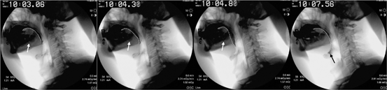
Fig. 9.1
Lateral fluoroscopic view demonstrates disordered oral preparatory phase. The oral tongue and tongue base (white dotted line) are pumping and there is bolus spillage into the oropharynx (black arrow) and the gingival buccal sulci (white arrow)
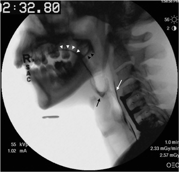
Fig. 9.2
Lateral fluoroscopic view demonstrates abnormal oral residue coating the oral tongue (white arrowheads) and palate (black arrowheads). Also seen is residue within the vallecula (black arrow) and pharyngoesophageal segment (white arrow). Additionally, the patient has anterior cervical spinal hardware in place
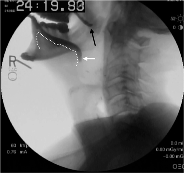
Fig. 9.3
Lateral fluoroscopic view of an individual who has had a near-total glossectomy for advanced head and neck cancer. A small remnant tongue is seen (white dotted line) as is a palatal prosthesis (black arrow). The patient has poor oral bolus control and early loss into the oropharynx (white arrow)
Table 9.2
Etiologies of oral tongue dysfunction
Etiology | Example |
|---|---|
Neoplasm/Posttreatment | Intrinsic lingual tumor Other oral cavity tumor CNS tumor External beam radiation Glossectomy |
Traumatic | Brain injury Facial trauma Hypoglossal nerve injury |
Neurologic | Stroke Parkinson’s disease Multiple sclerosis Cerebral palsy Muscular dystrophy Pseudobulbar palsy ALS |
Congenital | Ankyloglossia Macroglossia Trisomy 21 Pierre-Robin sequence |
Autoimmune/Inflammatory | Fungal infection Xerostomia Sjogren’s syndrome Myasthenia gravis Behcet’s disease |
Abnormal Pharyngeal Fluoroscopy
The junction of the hard and soft palates and circumvallate papillae at the anterior base of tongue delineate the beginning of the oropharynx . The oropharynx is critically important for safe and effective bolus propulsion and airway avoidance (Chap. 4). Waldeyer’s ring of lymphatic tissue includes the palatine, lingual, and pharyngeal tonsils, and is susceptible to pathologic enlargement secondary to acute infection, chronic inflammation, and neoplasm. Lymphatic abnormalities of Waldeyer’s ring can be visualized on VFSS (Fig. 9.4) and may result in dysfunction due to odynophagia, obstruction, or restricted base of tongue mobility. Hypertrophy of the lingual tonsil or base of tongue may be recognized on fluoroscopy as vallecular filing defects (Fig. 9.5). The vallecula may appear enlarged in individuals with base of tongue defects or atrophy (Fig. 9.6). Such findings may be due to neurologic processes, following surgical tongue base resection or radiation treatment to the pharynx. Physiologically, base of tongue deficiency may manifest as early bolus loss, incomplete oropharyngeal clearing and residue, or penetration and aspiration. Posteriorly, the vallecula is further bounded by the epiglottis, and irregularities of this structure are readily visualized on VFSS . Malformation, infection, and neoplasms may appear as a blurred, enlarged, or widened epiglottis (Fig. 9.7). Changes due to radiation therapy, surgery, or trauma may result in the appearance of a shortened or absent epiglottis.
Stay updated, free articles. Join our Telegram channel

Full access? Get Clinical Tree


