The Norwood Principle
Hypoplastic left heart syndrome is the most common form of congenital heart disease in which there is only one fully developed ventricle, and is the fourth most common congenital heart defect presenting in the first year of life. The anatomic features include aortic valve atresia or severe stenosis, with marked hypoplasia or absence of the left ventricle. The ascending aorta is small, usually only 2 to 3 mm in diameter, and the mitral valve is hypoplastic or atretic. A patent ductus arteriosus is the only route for adequate systemic perfusion.
Other single-ventricle complexes may present with evident or potential left ventricular outflow tract obstruction. These include tricuspid atresia with transposition of the great arteries and single ventricles with left ventricular outflow chambers. Narrowing of the bulboventricular foramen may cause subaortic obstruction. Pulmonary artery banding in this subgroup of patients may predispose to the development of subaortic obstruction.
The Norwood principle can be applied to all patients with single-ventricle morphology and real or potential obstruction to systemic flow. It involves the creation or preservation of optimal hemodynamics and anatomy in preparation for a successful Fontan procedure.
There are three important basic concepts in the initial palliation phase:
The aorta must be associated directly with the single ventricle in such a way as to provide unobstructed flow from the single ventricle to the systemic circulation and to allow potential for growth.
Pulmonary blood flow must be regulated to avoid the development of pulmonary vascular disease, and minimize the volume load on the single ventricle to preserve long-term ventricular function. This must be achieved without distorting the pulmonary arteries.
When there is stenosis or atresia of the left-sided atrioventricular valve, a large interatrial communication must be created to avoid the development of pulmonary venous obstruction and hypertension.
Stage I Palliative Reconstruction for Hypoplastic Left Heart Syndrome
Diagnosis may be made prenatally with a fetal echocardiogram, which then allows for prospective management of the neonate from the time of delivery. Preoperative management includes continuous infusion of prostaglandin E1 to maintain patency of the ductus arteriosus. A balanced pulmonary and systemic circulation is critical to the survival of these patients. A restrictive interatrial communication limits pulmonary overcirculation, and a balloon atrial septostomy or blade septectomy may result in hemodynamic deterioration and should be avoided. In addition, hyperventilation and increased inspired oxygen concentrations may decrease pulmonary vascular resistance, leading to increased pulmonary blood flow at the expense of decreased systemic perfusion. Hypoventilation with room air is most often indicated in these patients.
Traditionally, this operation has been performed with the use of deep hypothermic arrest for the aortic reconstruction. More recently, techniques using selective cerebral perfusion to avoid or minimize circulatory arrest have been adopted in most institutions.
Incision
A median sternotomy is performed, and the thymus is excised.
Cannulation
A purse-string suture of 6-0 Prolene is placed on the anterior wall of the main pulmonary artery, approximately 1 cm above the level of the valve, and on the right atrial appendage. The right and left pulmonary arteries are encircled with Silastic tapes. The arterial line is fitted with a Y-connector and two segments of tubing, one with a straight arterial cannula, and the other attached to a plastic olive-tipped catheter, which is clamped. The arterial cannula is deaired and introduced several millimeters into the pulmonary artery, and the purse-string suture is tightened. A single venous cannula is placed in the right atrial appendage and cardiopulmonary bypass is begun. The tapes around the pulmonary arteries are placed on traction to prevent pulmonary blood flow and ensure satisfactory systemic perfusion. During the cooling period, the ascending aorta is dissected away from the main pulmonary artery and the branch vessels of the aortic arch are mobilized. The innominate, left carotid, and left subclavian arteries are looped with Silastic tapes on
tourniquets. The distal aortic arch and descending aorta are mobilized down to the level of the left bronchus with blunt dissection. Regardless of the type of shunt ultimately planned for pulmonary blood flow, a 3.0- or 3.5-mm GORE-TEX tube graft is anastomosed to the innominate artery with a continuous 7-0 Prolene suture.
tourniquets. The distal aortic arch and descending aorta are mobilized down to the level of the left bronchus with blunt dissection. Regardless of the type of shunt ultimately planned for pulmonary blood flow, a 3.0- or 3.5-mm GORE-TEX tube graft is anastomosed to the innominate artery with a continuous 7-0 Prolene suture.
 Before cannulation, ice is placed around the patient’s head.
Procedure
After cooling for at least 10 to 15 minutes to a temperature of 18°C, the second limb of the arterial circuit is flushed and secured to the GORE-TEX tube. Alternatively, a single arterial line is used, and during a brief period of hypothermic arrest, the arterial cannula is removed from the pulmonary artery and placed into the tube graft. The previously placed tourniquets are used to occlude the proximal innominate artery, the left carotid, and left subclavian artery (Fig 30-1). The cannula in the pulmonary artery is removed. A curved clamp is placed on the distal descending aorta. The pump flow is reduced to 10 to 20 mL per kilogram per minute and adjusted to keep the right radial pressure between 30 and 40 mm Hg.
 In the presence of an anomalous right subclavian artery, the pressure in the right temporal artery must be monitored.
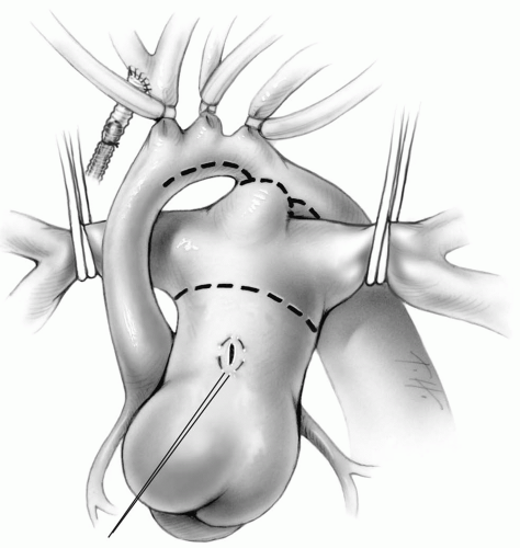 FIG 30-1. Occlusion of pulmonary arteries during cooling. GORE-TEX tube anastomosed to innominate artery for cerebral perfusion. Tourniquets are tightened around innominate, left carotid, and left subclavian arteries during low-flow cerebral perfusion. |
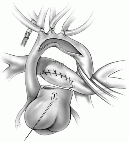 FIG 30-2. Transection of main pulmonary artery and patch closure of confluence. Oversewing of pulmonary end of ductus and opening of proximal descending aorta and arch. |
With the descending thoracic aorta and arch vessels occluded, cold blood cardioplegic solution is infused into the cannulation site of the main pulmonary artery. This perfuses the coronary circulation by retrograde flow through the ductus, arch, and ascending aorta.
A brief period of circulatory arrest or continued low-flow cerebral perfusion providing venous return with a pump sucker is used to excise the septum primum. This can be accomplished through the right atrial cannulation site by temporarily removing the venous cannula. Alternatively, a small right atriotomy is made and closed with a 6-0 Prolene suture after creating an adequate interatrial communication.
The ductus arteriosus is transected, and the pulmonary end is ligated or oversewn with a running 6-0 Prolene suture. The main pulmonary artery is transected at the level of the take-off of the right pulmonary artery (Fig. 30-1). The defect in the distal pulmonary artery is then closed with a small patch of pulmonary homograft to prevent stenosis of the confluence of the right and left pulmonary arteries (Fig. 30-2).
Aortic Arch Reconstruction
The technique for reconstruction of the aortic arch has changed over the years to avoid real or potential
complications. A direct anastomotic technique was initially employed by Norwood, but abandoned because of arch reobstruction and central pulmonary artery stenosis. The traditional technique uses a large pulmonary homograft patch to enlarge the proximal descending aorta, arch, and ascending aorta. However, concerns about lack of growth and calcification of the homograft patch prompted the reintroduction of an all autologous technique, with modifications for specific anatomic considerations.
complications. A direct anastomotic technique was initially employed by Norwood, but abandoned because of arch reobstruction and central pulmonary artery stenosis. The traditional technique uses a large pulmonary homograft patch to enlarge the proximal descending aorta, arch, and ascending aorta. However, concerns about lack of growth and calcification of the homograft patch prompted the reintroduction of an all autologous technique, with modifications for specific anatomic considerations.
Patch Reconstruction Technique
The aortic opening of the ductus arteriosus is extended distally for 10 to 15 mm on the medial aspect of the descending aorta. The opening is carried proximally along the lesser curvature of the aortic arch and down the left side of the ascending aorta. This incision should stop at the level of the transected proximal pulmonary artery.
All the ductal tissue in the aortic arch and descending aorta must be excised, including resection of the coarctation ridge, if present. Sewing to ductal tissue may result in bleeding from the suture line or even dehiscence of a portion of the anastomosis. In addition, residual ductal tissue may lead to late stenosis of the reconstructed arch. In some patients, this may require the excision of a circumferential portion of the periductal aorta. The descending aortic segment is then anastomosed to the posterolateral aspect of the distal arch opening with a running 7-0 Prolene suture. Both suture ends are secured before proceeding with the patch.
A triangular patch of pulmonary homograft is tailored to reconstruct the proximal descending aorta, aortic arch, and ascending aorta. The anastomosis of the pulmonary homograft to the aorta is started at the most distal extent of the incision on the descending aorta with a 7-0 Prolene double-armed suture. The posterior suture line is continued onto the ascending aorta, stopping 5 mm above the proximal extent of the incision. The anterior suture line is accomplished with the other needle, again ending the suture line 5 mm short of the proximal ascending aortic opening.
A patch cut from an adult-sized pulmonary homograft has a natural curved shape, which mimics the curve of the underside of the aortic arch. It is easy to handle, and has good hemostatic properties. However, there are availability and cost issues, as well as concerns regarding viral transmission and the generation of cytotoxic antibodies, which may limit transplant options. Some surgeons have advocated the use of bovine pericardium, using two pieces cut in a curved shape and sewn together along their concave aspect to create an appropriately shaped aortic arch patch. More recently, a segment of bovine vein opened longitudinally and trimmed, has been suggested as aortic arch patch material.
Alternating traction on the left carotid tourniquet and the innominate artery tourniquet improves the exposure for performing the posterior and anterior suture line on the underside of the aortic arch.
The main pulmonary artery is anastomosed to the ascending aorta, taking care not to distort the aortic root. This is accomplished with multiple interrupted 7-0 Prolene sutures to avoid “purse-stringing” of the opening into the aortic root. This interrupted suture line is carried up to meet the suture lines connecting the ascending aorta to the pulmonary homograft patch. The interrupted sutures are tied, and the continuous suture lines secured (Fig. 30-3). The pulmonary homograft patch and base of the pulmonary artery are pulled upward, and the patch trimmed to create an appropriate hood. The pulmonary base is sewn to the patch with a running 7-0 Prolene suture (Fig. 30-4).
The homograft patch must not be too large or left too long because it may compress the central pulmonary
artery. This is especially problematic if the original ascending aorta is larger than 3 to 4 mm in diameter. Pulmonary homograft tissue is fairly distensible, and this must be taken into consideration when fashioning the patch.
artery. This is especially problematic if the original ascending aorta is larger than 3 to 4 mm in diameter. Pulmonary homograft tissue is fairly distensible, and this must be taken into consideration when fashioning the patch.
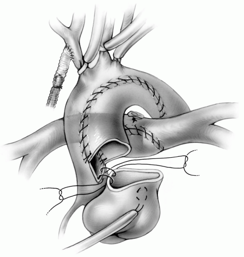 FIG 30-3. Reconstructing aorta with triangular patch of pulmonary homograft. Anastomosing proximal pulmonary artery to aortic root with interrupted sutures. |
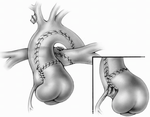 FIG 30-4. Completed aortic reconstruction with metal clip on innominate graft site. Inset: Distortion of proximal aorta by inaccurate alignment of proximal pulmonary artery to aortic opening or purse-stringing of anastomosis. |
Meticulous technique must be used when anastomosing a small ascending aorta to the proximal portion of the pulmonary artery to avoid obstructing flow into the coronary arteries (Fig. 30-4


Stay updated, free articles. Join our Telegram channel
Full access? Get Clinical Tree
