The Liver

Chapter 54 The Liver


Historical Perspective


The surface anatomy of the liver was described as early as 2000 years BC by the ancient Babylonians. Even Hippocrates understood and described the seriousness of liver injury. In 1654, Francis Glisson was the first physician to describe the essential anatomy of the blood vessels of the liver accurately. The beginnings of liver surgery are described as rudimentary excisions of eviscerated liver from penetrating trauma. The first documented case of a partial hepatectomy is credited to Berta, who amputated a portion of protruding liver in a patient with a self-inflicted stab wound in 1716.


In the late 1800s, the first gastrectomies and cholecystectomies were being performed in Europe. At that time, surgery on the liver was regarded as dangerous, if not impossible. In 1897, Elliot, in his report on liver surgery for trauma, said that the liver was so “friable, so full of gaping vessels and so evidently incapable of being sutured that it had always seemed impossible to successfully manage large wounds of its substance.” European surgeons began to experiment with techniques of elective liver surgery on animals in the late 1800s. The credit for the first elective liver resection is a matter of debate and many surgeons have been given credit, but it certainly occurred during this time period.


The early 1900s saw some small but significant advances in liver surgery. Techniques for suturing major hepatic vessels and the use of cautery for small vessels were applied and reported. The most significant advance of that time was probably that of J. Hogarth Pringle. In 1908, he described digital compression of the hilar vessels to control hepatic bleeding from traumatic injuries. The modern era of hepatic surgery was ushered in by the development of a better understanding of liver anatomy and formal anatomic liver resection. Credit for the first anatomic liver resection is usually given to Lortat-Jacob, who performed a right hepatectomy in 1952 in France. Pack from New York and Quattelbaum from Georgia performed similar operations within the next year and were unlikely to have had any knowledge of Lortat-Jacob’s report. Descriptions of the segmental nature of liver anatomy by Couinaud, Woodsmith, and Goldburne in 1957 opened the door even wider and introduced the modern era of liver surgery.


Despite these improvements, hepatic surgery was plagued by tremendous operative morbidity and mortality from the 1950s into the 1980s. Operative mortality rates in excess of 20% were common and usually related to massive hemorrhage. Many surgeons were reluctant to perform hepatic surgery because of these results and, understandably, many physicians were reluctant to refer patients for hepatectomy. With the courage of patients and their families, as well as the persistence of surgeons, safe hepatic surgery has now been realized. A complete list is not possible here, but courageous hepatic surgeons such as Blumgart, Bismuth, Longmire, Fortner, Schwartz, Starzl, and Ton deserve mention.


Advances in anesthesia, intensive care, antibiotics, and interventional radiologic techniques have also contributed tremendously to the safety of major hepatic surgery. Total hepatectomy with liver transplantation and live donor partial hepatectomy for transplantation are now performed routinely in specialized transplantation centers. Partial hepatectomy for a large number of indications is now performed throughout the world in specialized centers, with mortality rates of 5% or less. Partial hepatectomy performed on normal livers is now consistently performed, with mortality rates of 1% to 2%.


Safely performed open hepatic surgery and its liberal use in the management of a wide variety of diseases is now a reality. Moreover, minimally invasive approaches to liver surgery have been developed and are now being used in significant numbers. However, the learning curve remains steep and the indications for this technique are still being carefully defined. Thermal ablative techniques to treat hepatic tumors, including radiofrequency and microwave ablation, have exploded in popularity. Finally, techniques to improve the safety of liver resection further, such as portal vein embolization to induce preoperative hypertrophy of the future liver remnant (FLR), have been developed and are now being used.


Anatomy and Physiology


Anatomy


Gross Anatomy


A precise knowledge of the anatomy of the liver is an absolute prerequisite to performing surgery on the liver or biliary tree. With the development of hepatic surgery over the last several decades, a greater appreciation for the complex anatomy beyond the misleading minimal external markings has been realized. The days of using the falciform ligament as the only marker of the left and right sides of the liver are over; the anatomic contributions of Couinaud (see later) and the description of the segmental nature of the liver should be embraced and studied by students of hepatic surgery.


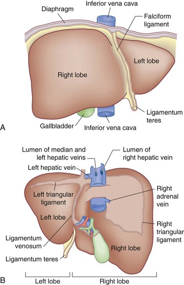
General Description and Topography


The liver is a solid gastrointestinal organ whose mass (1.2 to 1.6 kg) largely occupies the right upper quadrant of the abdomen. The costal margin coincides with the lower margin of the liver and the diaphragm drapes over the superior surface of the liver. The large majority of the right liver and most of the left liver is covered by the thoracic cage. The liver extends superiorly to the height of the fifth rib on the right and the sixth rib on the left. The posterior surface straddles the inferior vena cava (IVC). A wedge of liver extends to the left side of the abdomen. It crosses the epigastrium to lie above the anterior surface of the stomach and below the central and left portions of the diaphragm. The superior surface of the liver is convex and is molded to the diaphragm, whereas the inferior surface is mildly concave and extends to a sharp anterior border.


The liver is invested in peritoneum except for the gallbladder fossa, porta hepatis, and posterior aspect of the liver on either side of the IVC in two wedge-shaped areas. The region of liver to the right of the IVC is called the bare area of the liver.


The peritoneal duplications on the liver surface are referred to as ligaments. The diaphragmatic peritoneal duplications are referred to as the coronary ligaments whose lateral margins on either side are the right and left triangular ligaments. From the center of the coronary ligament emerges the falciform ligament, which extends anteriorly as a thin membrane connecting the liver surface to the diaphragm, abdominal wall, and umbilicus.


The ligamentum teres (the obliterated umbilical vein) runs along the inferior edge of the falciform ligament from the umbilicus to the umbilical fissure. The umbilical fissure is on the inferior surface of the left liver and contains the left portal pedicle. In early descriptions of hepatic anatomy, the falciform ligament, the most obvious surface marker of the liver, was used as the division of the right and left lobes of the liver. However, this description is inaccurate and of minimal usefulness to the hepatobiliary surgeon (see later for detailed segmental anatomy).


On the posterior surface of the left liver, running from the left portal vein in the porta hepatis toward the left hepatic vein and the IVC, is the ligamentum venosum (obliterated sinus venosus) which also runs in a fissure (Fig. 54-1). Hepatic arterial and portal venous blood enter the liver at the hilum and branch throughout the liver as a single portal pedicle unit, which also includes a bile duct. These portal triads are invested in a peritoneal sheath that invaginates at the hepatic hilum. Venous drainage is through the right, middle, and left hepatic veins that empty directly into the suprahepatic IVC.


Normal Development and Embryology


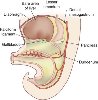
The developing liver shares a common progenitor with the biliary tree and pancreas. During embryogenesis, signals are transmitted from the cardiac mesenchyme and septum transversum. The molecules regulating this (e.g., FGF, BMP, Wnt) have begun to be elucidated. The liver primordium begins to form in the third week of development as an outgrowth of endodermal epithelium, known as the hepatic diverticulum or liver bud, known as the hepatic field. The connection between the hepatic diverticulum and the future duodenum narrows to form the bile duct and an outpouching of the bile duct forms into the gallbladder and cystic duct. Hepatic cells develop cords and intermingle with the vitelline and umbilical veins to form hepatic sinusoids. Simultaneously, hematopoietic cells, Kupffer cells and connective tissue form from the mesoderm of the septum transversum. The mesoderm of the septum transversum connects the liver to the ventral abdominal wall and foregut. As the liver protrudes into the abdominal cavity, these structures are stretched into thin membranes, ultimately forming the falciform ligament and lesser omentum, respectively. The mesoderm on the surface of the developing liver differentiates into visceral peritoneum, except superiorly, where contact between the liver and mesoderm (future diaphragm) is maintained, forming a bare area devoid of visceral peritoneum (Fig. 54-2).


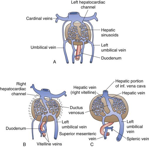
The primitive liver plays a central role in the fetal circulation. The vitelline veins carry blood from the yolk sac to the sinus venosus and ultimately form a network of veins around the foregut (future duodenum) that drain into the developing hepatic sinusoids. These vitelline veins eventually fuse to form the portal, superior mesenteric, and splenic veins. The sinus venosus, which empties into the fetal heart, becomes the hepatocardiac channel and then the hepatic veins and retrohepatic IVC. The umbilical veins, which are paired early on, carry oxygenated blood to the fetus. Initially, the umbilical veins drain into the sinus venosus but at week 5 of development, they begin to drain into the hepatic sinusoids. The right umbilical vein ultimately disappears and the left umbilical vein later drains directly into the hepatocardiac channel, bypassing the hepatic sinusoids through the ductus venosus. In the adult liver, the remnant of the left umbilical vein becomes the ligamentum teres, which runs in the falciform ligament into the umbilical fissure, and the remnant of the ductus venosus becomes the ligamentum venosus at the termination of the lesser omentum under the left liver (Fig. 54-3).


The fetal liver plays a very important role in hematopoiesis. In week 10 of gestation, the liver comprises 10% of the fetal body weight because of developing hepatic sinusoids and active hematopoiesis. During the last 2 months of intrauterine life, hepatic hematopoiesis decreases and the weight of the liver is decreased to 5% of the fetal body weight.


By week 12 of gestation, bile is formed in hepatocytes and secreted into the bile ductules of each hepatic lobule. Simultaneously, bile duct epithelial cells (cholangiocytes) develop along intrahepatic and extrahepatic bile ducts while the gallbladder completes its development. Together, this allows for the drainage of bile into the foregut.


The adult liver is a complex system of numerous cell types, including hepatocytes, cholangiocytes, neuroendocrine cells, hepatic progenitors (known as oval cells), myofibroblastic mesenchymal cells (known as hepatic stellate cells and portal myofibroblasts), resident macrophages (known as Kupffer cells), and vascular endothelial cells.


Functional Anatomy


Historically, the liver was divided into left and right lobes by the obvious external landmark of the falciform ligament. Not only was this description oversimplified, but it was anatomically incorrect in relation to the blood supply to the liver. Later, a more accurate understanding of the lobar anatomy of the liver was developed. The liver is divided into right and left lobes determined by portal and hepatic vein branches.


Our understanding of functional liver anatomy has become more sophisticated. Briefly, a plane without any surface markings, known as the portal fissure or Cantlie’s line, runs from the gallbladder to the left side of the IVC. This divides the liver into right and left lobes. The right lobe is further divided into anterior and posterior sectors. The left lobe is divided into a medial sector, also known as the quadrate lobe, that lies to the right of the falciform ligament and umbilical fissure and a lateral sector, also known as the left lateral segment, which lies to the left of these structures. This system, although anatomically more correct, is only sufficient for mobilization of the liver and simple hepatic resections. It does not describe the more intricate and functional segmental anatomy that is essential to understand before pursuing complex hepatobiliary surgery.


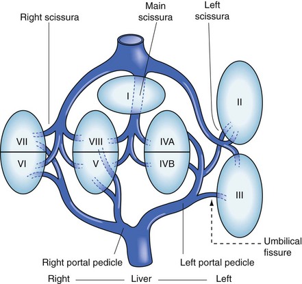
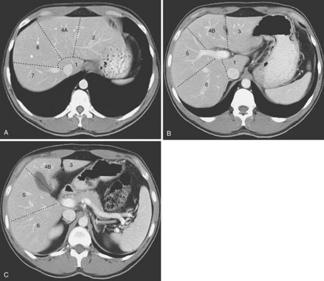
The functional anatomy of the liver (Figs. 54-4 and 54-5) is composed of eight segments, each supplied by a single portal triad (also called a pedicle) composed of a portal vein, hepatic artery, and bile duct. These segments are further organized into four sectors separated by scissurae containing the three main hepatic veins. The four sectors are even further organized into the right and left liver. The terms right liver and left liver are preferable to the terms right lobe and left lobe because there is no external mark that allows the identification of the right and left liver. This system was originally described in 1957 by Woodsmith and Goldburne, and by Couinaud. It defines hepatic anatomy because it is most relevant to surgery of the liver. The functional anatomy is more often seen as cross-sectional imaging (Fig. 54-6).


The main scissura contains the middle hepatic vein, which runs in an anteroposterior direction from the gallbladder fossa to the left side of the vena cava. It divides the liver into right and left hemilivers. The line of the main scissura is also known as Cantlie’s line (see earlier). The right liver is divided into anterior (segments 5 and 8) and posterior (segments 6 and 7) sectors by the right scissura, which contains the right hepatic vein. The right portal pedicle is composed of the right hepatic artery, portal vein, and bile duct. It splits into right anterior and right posterior pedicles, which supply the segments of the anterior and posterior sectors.


The left liver has a visible fissure along its inferior surface called the umbilical fissure. The ligamentum teres, containing the remnant of the umbilical vein, runs into this fissure. The falciform ligament is contiguous with the umbilical fissure and ligamentum teres. The umbilical fissure is not a scissura and does not contain a hepatic vein; it contains the left portal pedicle, which contains the left portal vein, hepatic artery, and bile duct. This pedicle runs in this fissure and branches to feed the left liver. The left liver is split into anterior (segments 3 and 4) and posterior (segment 2, the only sector composed of a single segment) sectors by the left scissura. The left scissura runs posterior to the ligamentum teres and contains the left hepatic vein.


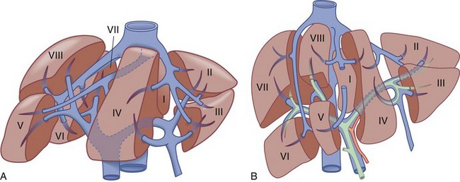
At the hilum of the liver, the right portal triad has a short extrahepatic course of approximately 1 to 1.5 cm before entering the substance of the liver and branching into anterior and posterior sectoral branches. The left portal triad, however, has a long extrahepatic course of up to 3 to 4 cm and runs transversely along the base of segment 4 in a peritoneal sheath, which is the upper end of the lesser omentum. This connective tissue is known as the hilar plate (Fig. 54-7). The continuation of the left portal triad runs anteriorly and caudally in the umbilical fissure and gives branches to segments 2 and 3 and recurrent branches to segment 4.


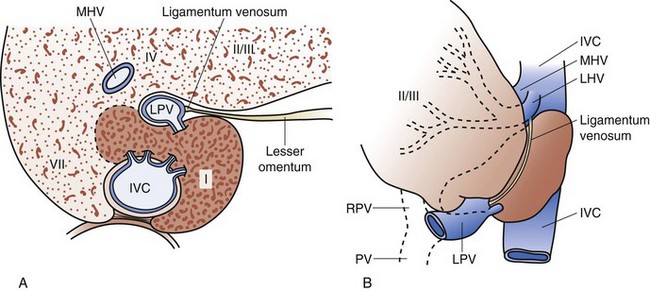
The caudate lobe (segment 1) is the dorsal portion of the liver. It embraces the IVC on its posterior surface and lies posterior to the left portal triad inferiorly and the left and middle hepatic veins superiorly. The main bulk of the caudate lobe is to the left of the IVC, but inferiorly it traverses between the IVC and left portal triad, where it fuses to the right liver (segments 6 and 7). This part of the caudate lobe is known as the right portion or the caudate process. The left portion of the caudate lobe lies in the lesser omental bursa and is covered anteriorly by the gastrohepatic ligament (lesser omentum) that separates it from segments 2 and 3 anteriorly. The gastrohepatic ligament attaches to the ligamentum venosum (sinus venosus remnant) along the left side of the left portal triad (Fig. 54-8).
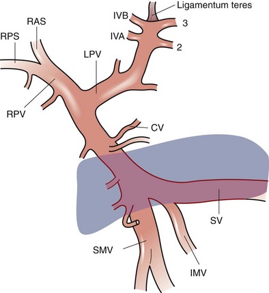


The vascular inflow and biliary drainage to the caudate lobe comes from both the right and left pedicles. The right side of the caudate, the caudate process, largely derives its portal venous supply from the right portal vein or the bifurcation of the main portal vein. The left portion of the caudate derives its portal venous inflow from the left main portal vein. The arterial supply and biliary drainage of the right portion are generally through the right posterior pedicle system and the left portion through the left main pedicle. The hepatic venous drainage of the caudate is unique because a number of posterior small veins drain directly into the IVC.


The posterior edge of the left side of the caudate terminates as a fibrous component that attaches to the crura of the diaphragm and also runs posteriorly, wrapping behind the IVC and attaching to segment 7 of the right liver. In up to 50% of people, this fibrous component is composed partially or completely of liver parenchyma. Thus, liver tissue may completely encircle the IVC. This structure is known as the caval ligament and is important to recognize when mobilizing the right liver or the caudate lobe off the vena cava.


Anomalous development of the liver is uncommonly encountered. Complete absence of the left liver has been reported. A tongue of tissue extending inferiorly off the right liver has been described (Riedel’s lobe). Rare cases of supradiaphragmatic liver in the absence of a hernia sac have been noted.


Portal Vein


The portal vein provides approximately 75% of the hepatic blood inflow. Despite being postcapillary and largely deoxygenated, its high flow rate provides 50% to 70% of the liver’s oxygen. The lack of valves in the portal venous system provides a system that can accommodate high flow at low pressure because of the low resistance. This allows for the measurement of portal venous pressure at any point along the system.


The portal vein forms behind the neck of the pancreas at the confluence of the superior mesenteric vein and the splenic vein at the level of the second lumbar vertebrae. The length of the main portal vein ranges from 5.5 to 8 cm and its diameter is usually approximately 1 cm. Cephalad to its formation behind the neck of the pancreas, the portal vein runs behind the first portion of the duodenum and into the hepatoduodenal ligament, where it runs along the right border of the lesser omentum, usually posterior to the common bile duct and proper hepatic artery.


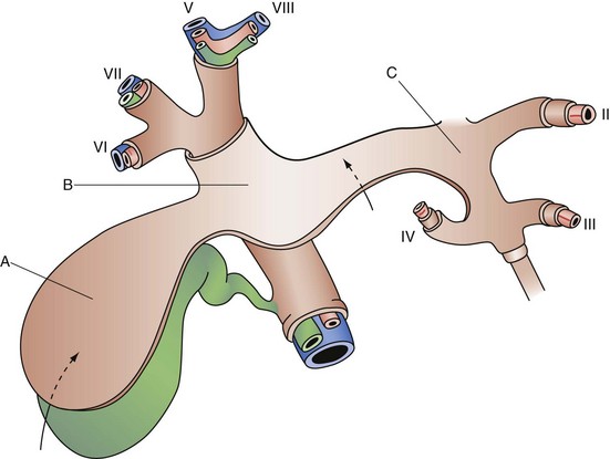
The portal vein divides into main right and left branches at the hilum of the liver. The left branch of the portal vein runs transversely along the base of segment 4 and into the umbilical fissure, where it gives off branches to segments 2 and 3 and feedback branches to segment 4. The left portal vein also gives off posterior branches to the left side of the caudate lobe. The right portal vein has a short extrahepatic course; it usually enters the substance of the liver where it splits into anterior and posterior sectoral branches. These sectoral branches can occasionally be seen extrahepatically and can come off the main portal vein before its bifurcation. There is usually a small caudate process branch off the main right portal vein or at the right portal vein bifurcation, which comes off posteriorly to supply this portion of liver (Fig. 54-9).


There are a number of connections between the portal and systemic venous systems. Under conditions of high portal venous pressure, these portosystemic connections may enlarge secondarily to collateral flow. This concept is reviewed in more detail in later in the chapter, but the most significant portosystemic collateral locations are the following: (1) the submucosal veins of the proximal stomach and distal esophagus receive portal flow from the short gastric veins and the left gastric vein and can result in varices, with the potential for hemorrhage; (2) the umbilical and abdominal wall veins recanalize from flow through the umbilical vein in the ligamentum teres, resulting in caput medusae; (3) the superior hemorrhoidal plexus receives portal flow from inferior mesenteric vein tributaries and can form large hemorrhoids; and (4) other retroperitoneal communications yield collaterals that can make abdominal surgery hazardous.


The anatomy of the portal vein and its branches is relatively constant and has much less variation than the biliary ductal and hepatic arterial systems. The portal vein is rarely found anterior to the neck of the pancreas and duodenum. Entrance of the portal vein directly into the vena cava has also been described. Very rarely, a pulmonary vein may enter the portal vein. Finally, there may be a congenital absence of the left branch of the portal vein. In this situation, the right branch courses through the right liver and curves around peripherally to supply the left liver, or the right anterior sectoral vein can arise from the left portal vein.


Hepatic Artery


The hepatic artery, representing high-volume oxygenated systemic arterial flow, provides approximately 25% of the hepatic blood flow and 30% to 50% of its oxygenation. A number of smaller perihepatic arteries derived from the inferior phrenic and the gastroduodenal arteries also supply the liver. These vessels are important sources of collateral blood flow in case of occlusion of the main hepatic arterial inflow. In the case of ligation of the right or left hepatic artery, intrahepatic collaterals almost immediately provide for nutrient blood flow in most cases.


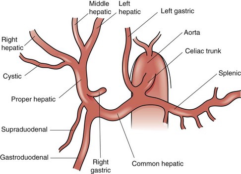
The common description of the arterial supply to the liver and biliary tree is only present approximately 60% of the time (Fig. 54-10). The celiac trunk originates directly off the aorta, just below the aortic diaphragmatic hiatus, and gives off three branches—the splenic artery, left gastric artery, and common hepatic artery. The common hepatic artery passes forward and to the right along the superior border of the pancreas and runs along the right side of the lesser omentum, where it ascends towards the hepatic hilum, lying anterior to the portal vein and to the left of the bile duct. At the point where the common hepatic artery begins to head superiorly towards the hepatic hilum, it gives off the gastroduodenal artery, followed by the supraduodenal artery and right gastric artery. The common hepatic artery beyond the takeoff of the gastroduodenal is called the proper hepatic artery; it divides into right and left hepatic arteries at the hilum. The left hepatic artery heads vertically towards the umbilical fissure to supply segments 2, 3, and 4. The left hepatic artery usually also gives off a middle hepatic artery branch that heads toward the right side of the umbilical fissure and supplies segment 4. The right hepatic artery usually runs posterior to the common hepatic bile duct and enters Calot’s triangle, bordered by the cystic duct, common hepatic duct, and liver edge, where it gives off the cystic artery to supply the gallbladder and then continues into the substance of the right liver.


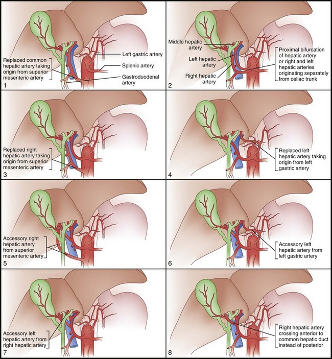
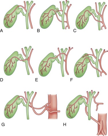
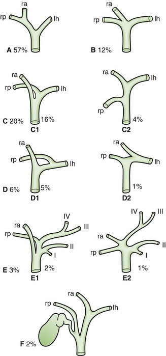
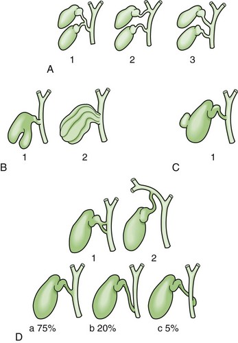
Unlike portal vein anatomy, hepatic arterial anatomy is extraordinarily variable (Fig. 54-11). An accessory vessel is described as an aberrant origin of a branch that is in addition to the normal branching pattern. A replaced vessel is described as an aberrant origin of a branch that substitutes for the lack of the normal branch. Usually, the hepatic artery originates off the celiac trunk. However, branches or the entire hepatic arterial system can originate off the superior mesenteric artery (SMA). The right and left hepatic arteries can also arise separately off the celiac axis. Replaced or accessory right hepatic arteries come off the SMA and are present approximately 11% to 21% of the time. Hepatic vessels replaced to the SMA run behind the head of the pancreas, posterior to the portal vein in the portacaval space. The right hepatic artery, in its usual branching pattern, can also course anterior to the common hepatic duct. A replaced or accessory left hepatic artery is present approximately 3.8% to 10% of the time, originates from the left gastric artery, and courses within the lesser omentum, heading toward the umbilical fissure. Other important variations include the origin of the gastroduodenal artery, which has been found to originate from the right hepatic artery and is occasionally duplicated. The anatomy of the cystic artery is also variable; knowledge of these variations is of particular importance in the performance of cholecystectomy (Fig. 54-12). An accessory cystic artery can originate from the proper hepatic artery or gastroduodenal artery, where it runs anterior to the bile duct. A single cystic artery can originate anywhere off the proper hepatic artery or gastroduodenal artery, or directly from the celiac axis. These variant cystic arteries can run anterior to the bile duct and are not necessarily present in the triangle of Calot. All these variations in hepatic arterial anatomy are of obvious importance during hepatic resection, hepatic arterial pump placement, cholecystectomy, and hepatic interventional radiologic procedures.


Hepatic Veins


The three major hepatic veins drain from the superior-posterior surface of the liver directly into the IVC (see Figs. 54-4, 54-5, and 54-6). The right hepatic vein runs in the right scissura between the anterior and posterior sectors of the right liver and drains most of the right liver after a short (1-cm) extrahepatic course into the right side of the IVC. The left and middle hepatic veins usually join intrahepatically and enter the left side of the IVC as a single vessel, although they may drain separately. The left hepatic vein runs in the left scissura between segments 2 and 3 and drains segments 2 and 3; the middle hepatic vein runs in the portal scissura between segment 4 and the anterior sector of the right liver, comprised of segments 5 and 8, and drains segment 4 and some of the anterior sector of the right liver. The umbilical vein is an additional vein that runs under the falciform ligament, between the left and middle veins, and usually empties into the left hepatic vein. A number of small posterior venous branches from the right posterior sector and caudate lobe drain directly into the IVC. A substantial inferiorly located accessory right hepatic vein is commonly encountered. There is also often a venous tributary from the caudate lobe, which drains superiorly into the left hepatic vein.


Biliary System


The intrahepatic bile ducts are the terminal branches of the right and left hepatic ductal branches that invaginate Glisson’s capsule at the hilum, along with their corresponding portal vein and hepatic artery branches, forming the peritoneal covered portal triads also known as portal pedicles. Along these intrahepatic portal pedicles, the bile duct branches are usually superior to the portal vein, whereas the hepatic artery branches run inferiorly. The left hepatic bile duct drains segments 2, 3, and 4, which constitute the left liver. The intrahepatic ductal branches of the left liver join to form the main left duct at the base of the umbilical fissure, where the left hepatic duct courses transversely across the base of segment 4 to join the right hepatic duct at the hilum. In its transverse portion, the left hepatic duct drains one to three small branches from segment 4. The right hepatic duct drains the right liver and is formed by the joining of the anterior sectoral duct (draining segments 5 and 8) and the posterior sectoral duct (draining segments 6 and 7). The posterior sectoral duct runs in a horizontal and posterior direction; the anterior sectoral duct runs vertically. The main right hepatic duct bifurcates just above the right portal vein. The short right hepatic duct meets the longer left hepatic duct to form the confluence anterior to the right portal vein, constituting the common hepatic duct. The caudate lobe (segment 1) has its own biliary drainage, which is usually through right and left systems. However, in up to 15% of individuals, drainage is through the left system only and, in 5%, it is through the right system only.


The common hepatic duct drains inferiorly. Below the takeoff of the cystic duct, it is referred to as the common bile duct. The common bile duct usually measures 10 to 15 cm in length and is typically 6 mm in diameter. The common hepatic (bile) duct runs along the right side of the hepatoduodenal ligament (free edge of the lesser omentum) to the right of the hepatic artery and anterior to the portal vein. The common bile duct continues inferiorly behind the first portion of the duodenum and into the head of the pancreas in an inferior and slightly rightward direction. The intrapancreatic distal common bile duct then joins with the main pancreatic duct (of Wirsung), with or without a common channel, and enters the second portion of the duodenum through the major papilla of Vater. At the choledochoduodenal junction, a complex muscular complex known as the sphincter of Oddi regulates bile flow and prevents reflux of duodenal contents into the biliary tree. There are three major parts to this sphincter: (1) the sphincter choledochus, which is a circular muscle that regulates bile flow and the filling of the gallbladder; (2) the pancreatic sphincter, present to variable degrees, which surrounds the intraduodenal pancreatic duct; and (3) the sphincter ampullae, made up of longitudinal muscle, which prevents duodenal reflux.


The gallbladder is a biliary reservoir that lies against the inferior surface of segments 4 and 5 of the liver, usually making an impression against the liver. A peritoneal layer covers most of the gallbladder, except for the portion adherent to the liver. Here, the gallbladder adheres to the liver by a layer of fibroconnective tissue known as the cystic plate, an extension of the hilar plate (see Fig. 54-7). Variable in size, but usually about 10 cm long and 3 to 5 cm wide, the gallbladder is composed of a fundus, body, infundibulum, and neck, which ultimately empty into the cystic duct. The fundus usually projects just slightly beyond the liver edge anteriorly and, when folded on itself, is described as a Phrygian cap. Continuing toward the bile duct, the body of the gallbladder is usually in close proximity to the second portion of the duodenum and transverse colon. The infundibulum (or Hartmann’s pouch) hangs forward along the free edge of the lesser omentum and can fold in front of the cystic duct. The portion of gallbladder between the infundibulum and cystic duct is referred to as the neck. The cystic duct is variable in its length, course, and insertion into the main biliary tree. The first portion of the cystic duct is usually tortuous and contains mucosal duplications, referred to as the folds of Heister, which regulate the filling and emptying of the gallbladder. Usually, the cystic duct joins the common hepatic duct to form the common bile duct.


Knowledge of the multiple and frequent variations in the anatomy of the biliary tree is absolutely essential for performing hepatobiliary procedures. Anomalies of the hepatic ductal confluence are common and are present approximately one third of the time. The most common anomalies of the biliary confluence involve variations in the insertion of the right sectoral ducts. Usually, this is the posterior sectoral duct. The confluence can be a trifurcation of the right anterior sectoral, right posterior sectoral, and left hepatic ducts. Either of the right sectoral ducts can drain into the left hepatic duct, common hepatic duct, cystic duct or, rarely, the gallbladder (Fig. 54-13).


Anomalies of the gallbladder itself are rare. Agenesis of the gallbladder, bilobar gallbladder with two ducts or a single duct, septations, and congenital diverticulum of the gallbladder have all been described. Anomalies of the position of the gallbladder are more common; these include an intrahepatic position or, rarely, located on the left side of the liver. The gallbladder can also have a long mesentery, which can predispose it to torsion.


The position and entry of the cystic duct into the main ductal system are also variable. Double cystic ducts draining a unilocular gallbladder and drainage into hepatic duct branches have been reported. Usually, the cystic duct joins the common hepatic duct at an angle, but can run parallel and enter it more distally. In the latter situation, the cystic duct can be fused to the hepatic duct along its parallel course by connective tissue. The cystic duct can also run a spiral course anteriorly or posteriorly and enter the left side of the common hepatic duct. Finally, the cystic duct can be very short or even absent (Fig. 54-14).


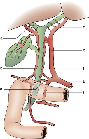
The supraduodenal and infrahilar bile duct are predominantly supplied by two axial vessels that run at 3- and 9-o’clock positions. These vessels are derived from the superior pancreaticoduodenal, right hepatic, cystic, gastroduodenal, and retroduodenal arteries. It has been estimated that only 2% of the arterial supply to this portion of the bile duct is segmental, arising directly off the proper hepatic artery. The bile duct and its bifurcation in the hilum derive their arterial blood supply from a rich network of multiple small branches from surrounding vessels. Similarly, the retropancreatic bile duct derives its arterial supply from the retroduodenal artery, which provides a rich network of multiple small branches (Fig. 54-15). Venous drainage of the bile duct parallels the arterial supply and drains into the portal venous system. The venous drainage of the gallbladder empties into the veins that drain the bile duct and does not flow directly into the portal vein.


Microscopic Anatomy


Functional Unit of the Liver


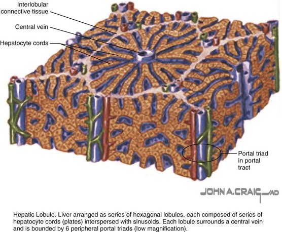
The organization of hepatic parenchyma into microscopic functional units has been described in a number of ways, referred to as an acinus or a lobule (Fig. 54-16). This was originally described by Rappaport and then modified by Matsumoto and Kawakami.6 A lobule is made up of a central terminal hepatic venule surrounded by four to six terminal portal triads that form a polygonal unit. This unit is lined on its periphery between each terminal portal triad by terminal portal triad branches. In between the terminal portal triads and the central hepatic venule, hepatocytes are arranged in one cell–thick plates, surrounded on each side by endothelial-lined and blood-filled sinusoids. Blood flows from the terminal portal triad through the sinusoids into the terminal hepatic venule. Bile is formed within the hepatocytes and empties into terminal canaliculi, which form on the lateral walls of the intercellular hepatocyte. These ultimately coalesce into bile ducts and flow toward the portal triads. This functional hepatic unit provides a structural basis for the many metabolic and secretory functions of the liver.


Between the terminal portal triad and central hepatic venule are three zones that differ in their enzymatic makeup, as well as exposure to nutrients and oxygenated blood. There is debate about the shape of these zones and their relationship to the basic lobular unit but, in general, zones 1 through 3 splay out from the terminal portal triad toward the central hepatic venule. Zone 1 (periportal zone) is an environment rich in nutrients and oxygen. Zones 2 (intermediate zone) and 3 (perivenular zone) are exposed to environments that are poorer in oxygen and nutrients. The cells of the different zones differ enzymatically and respond differently to toxin exposure and hypoxia. This anatomic arrangement also explains the phenomenon of centrilobular necrosis from hypotension, because zone 3 is the most susceptible to decreases in oxygen delivery.


Hepatic Microcirculation


Terminal portal venous and hepatic arterial branches directly supply the hepatic sinusoids with blood. The portal branches provide a constant-, but minimal flow into this low-volume system; the arterial branches provide the sinusoids with pulsatile-, but low-volume flow that enhances flow in the sinusoids. Hepatic arterial branches terminate in a plexus around the terminal bile ductules and provide nutrients. Arterial and portal vein flow vary inversely in the sinusoids and can be compensatory. Local control of blood flow in the sinusoids likely depends on arteriolar sphincters and contraction of the sinusoidal lining by endothelial cells and hepatic stellate cells or portal myofibroblasts. Blood within the sinusoids empties directly into terminal hepatic venules at the center of a functional lobule. This process results in the unidirectional flow of blood in the liver from zones 1 to 3.


The endothelium-lined sinusoids of the hepatic lobule represent the functional unit of the liver, where afferent blood flow is exposed to functional hepatic parenchyma prior to being drained into hepatic venules (Fig. 54-17). The hepatic sinusoids are 7 to 15 µm wide but can increase in size by up to 10-fold. This yields a low-resistance and low-pressure (generally, 2 to 3 mm Hg) system. The sinusoidal endothelial cells account for 15% to 20% of the total hepatic cell mass.
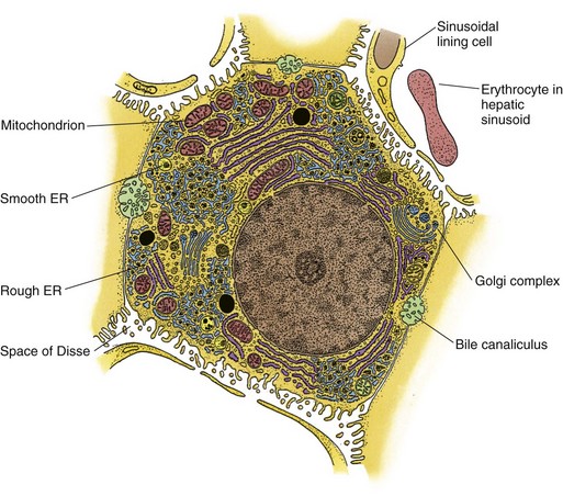


Sinusoidal endothelial cells are separated from hepatocytes by the space of Disse (perisinusoidal space). This is an extravascular fluid compartment into which hepatocytes project microvilli, which allows proteins and other plasma components from the sinusoids to be taken up by the hepatocytes. Within this space, the endothelial cells are specialized in that they lack intercellular junctions and a basement membrane but contain multiple large fenestrations. This arrangement provides for the maximal contact of hepatocyte membranes with this extravascular fluid compartment and blood in the sinusoidal space. Thus, this system permits bidirectional movement of solutes (high- and low-molecular-weight substances) into and out of hepatocytes, providing tremendous filtration potential. On the other hand, the fenestrations of the endothelial cells restrict movement of molecules between the sinusoids and hepatocytes and vary in response to exogenous and endogenous mediators.


Other cell types are found along the sinusoidal lining. Kupffer cells, derived from the macrophage-monocyte system, are irregularly-shaped cells that also line the sinusoids insinuating between endothelial cells. Kupffer cells are phagocytic, can migrate along sinusoids to areas of injury, and play a major role in the trapping of foreign substances and initiating inflammatory responses. Major histocompatibility complex II antigens are expressed on Kupffer cells, but do not confer efficient antigen presentation compared with macrophages elsewhere in the body. Other lymphoid cells also exist in hepatic parenchyma, such as natural killer (NK), natural killer T (NKT), CD4 T, and CD8 T cells. These provide the liver with an innate immune system. Hepatic stellate cells, previously known as Ito cells, are cells high in retinoid content (accounting for their phenotypic identification) found in the space of Disse. They have dendritic processes that contact hepatocyte microvilli and also wrap around endothelial cells. The major functions of these stellate cells include vitamin A storage and the synthesis of extracellular collagen and other extracellular matrix proteins. In acute and chronic hepatic liver injuries, hepatic stellate cells are activated to a myofibroblastic state associated with morphologic changes, cellular contractility, decreases in intracellular vitamin A, and production of extracellular matrix. Ultimately, stellate cells play a central role in the development and progression of hepatic fibrosis to cirrhosis and are the target for the development of antifibrotic treatments.


Hepatocytes


Hepatocytes are complex multifunctional cells that make up 60% of the hepatic cellular mass and 80% of the cytoplasmic mass of the liver (see Fig. 54-17). Morphologically, the hepatocyte is a polyhedral cell with a central spherical nucleus. As noted, hepatocytes are arranged in single cell layer plates lined on either side by blood-filled sinusoids. Every hepatocyte has contact with adjacent hepatocytes, the biliary space (bile canaliculus), and the perisinusoidal space, enabling these cells to perform their broad range of functions. Among the many essential functions of the hepatocyte are the following: (1) uptake, storage and release of nutrients; (2) synthesis of glucose, fatty acids, lipids, and numerous plasma protein (including C-reactive protein and albumin); (3) production and secretion of bile for digestion of dietary fats; and (4) degradation and detoxification of toxins.


To carry out these functions, the plasma membrane of the hepatocyte is organized in a specific manner into three specific domains. The sinusoidal membrane is exposed to the space of Disse and has multiple microvilli that provide a surface specialized in the active transport of substances between the blood and hepatocytes. The lateral domain exists between neighboring hepatocytes and contains gap junctions that provide for intercellular communication. The canalicular membrane is a tube containing microvilli formed by two apposed hepatocytes. These bile canaliculi are sealed by zonula occludens (tight junctions), which prevent the escape of bile. The bile canaliculi form a ring around the hepatocyte that drains into small bile ducts known as canals of Hering, which empty into a bile duct at a portal triad. The canalicular membrane contains adenosine triphosphate (ATP)–dependent active transport systems that enable solutes to be secreted into the canalicular membrane against large concentration gradients.


The hepatocyte is one of the most diverse and metabolically active cells in the body, as reflected by its abundance of organelles. There are 1000 mitochondria/hepatocyte, occupying approximately 20% of the cell volume. Mitochondria generate energy (ATP) through oxidative phosphorylation and provide the energy for the metabolic demands of the hepatocyte. The hepatocyte mitochondria are also essential for fatty acid oxidation. The monoclonal antibody HepPar1 (hepatocyte paraffin-1) identifies a unique antigen on hepatocyte mitochondria and is widely used to identify hepatocytes or hepatocellular neoplasms on immunohistochemical examination.


An extensive system of interconnected membrane complexes made up of smooth and rough endoplasmic reticulum and the Golgi apparatus comprise what is known as the hepatocyte microsomal fraction. These complexes have a diverse range of functions, including the following: (1) synthesis of structural and secreted proteins; (2) metabolism of lipids and glucose; (3) production and metabolism of cholesterol; (4) glycosylation of secretory proteins; (5) bile formation and secretion; and (6) drug metabolism. Finally, hepatocytes also contain lysosomes, which are intracellular single-membrane vesicles that contain a number of enzymes. These vesicles store and degrade exogenous and endogenous substances. Coordination of these numerous organelles in the hepatocyte allows these cells to accomplish a large variety of functions.


Functions


The unique anatomic arrangement of the liver described provides a remarkable landscape on which the multiple central and critical functions of this organ can be carried out. The liver is the center of metabolic homeostasis; it serves as the regulatory site for energy metabolism by coordinating the uptake, processing, and distribution of nutrients and their subsequent energy products. The liver also synthesizes a large number of proteins, enzymes, and vitamins that participate in a tremendously broad range of bodily functions. Finally, the liver detoxifies and eliminates many exogenous and endogenous substances, serving as the major filter of the human body. The following sections will summarize this broad range of functions.


Energy


The liver is the critical intermediary between dietary sources of energy and the extrahepatic tissues that require this energy. The critical and central nature of the liver in regulating the body’s energy metabolism is evidenced by the fact that despite accounting for only 4% of the total body weight, the liver consumes about 28% of the total body blood flow and 20% of the total oxygen consumed. The liver also uses about 20% of the total body caloric intake.


The liver receives dietary byproducts through the portal circulation and sorts, metabolizes, and distributes them into the systemic circulation. The liver also plays a major role in regulating endogenous sources of energy such as fatty acids and glycerol from adipose tissues and lactate, pyruvate, and certain amino acids from skeletal muscle. The two major sources of energy that the liver releases into the extrahepatic circulation are glucose and acetoacetate. Glucose is derived from the glycogenolysis of stored glycogen and from gluconeogenesis from lactate, pyruvate, glycerol, propionate, and alanine. Acetoacetate is derived from the β-oxidation of fatty acids. Also, storage lipids such as triacylglycerols and phospholipids are synthesized and stored as lipoproteins by the liver. These can be circulated systemically for uptake by peripheral tissues. These complex and essential functions are regulated by hormones, overall nutritional state of the organism, and requirements of obligate glucose-requiring tissues.


Functional Heterogeneity


To add to the metabolic complexity of the liver, hepatocytes vary in their function, depending on their location within the lobule. This functional heterogeneity of hepatocytes is anatomically related to their location in the three zones of the lobule and is specifically related to the distance from the incoming portal triad. For example, cells located in the periportal zone (zone 1) are exposed to a high concentration of substrates. Thus, uptake of oxygen and solutes are greater here. A critically important function of hepatocytes, however, is their ability to change their metabolic functionality and be recruited to perform specific functions under varying physiologic conditions, regardless of anatomic location. Sinusoids in the periportal zone are narrower and more tortuous, facilitating increased uptake of substrate by the hepatocytes in this area. In contrast, sinusoids in zone 3 (perivenous) have larger fenestrations, allowing uptake of larger molecules. Thus, sinusoids are also variable in form and function.


Enzymatic makeup, plasma membrane proteins, and ultrastructure are also heterogenous among the hepatocyte population. This cellular protein variability can also be distinguished based on the hepatocyte location within the lobule. Glucose uptake and release, bile formation, and synthesis of albumin and fibrinogen take place in the periportal zone, whereas glucose catabolism, xenobiotic metabolism, and synthesis of α1-antitrypsin and α-fetoprotein (AFP) occur in the perivenous zone. Another example of enzymatic heterogeneity according to lobular zones is the location of the urea cycle enzymes in zone 3, adjacent to the terminal hepatic veins. The functional hepatocyte heterogeneity and its anatomic relationship to the lobular unit account for patterns of damage from metabolic or physiologic insults to the liver.


Blood Flow


There is a dual blood supply to the liver that comes from the portal vein and hepatic artery. The portal vein provides approximately 75% of the blood flow to the liver, which is oxygen-poor but rich in nutrients. The hepatic artery provides the other 25% of the blood flow, which is oxygen- rich and represents systemic arterial blood flow. The large flow rate of the portal vein is still able to provide 50% to 70% of the afferent oxygenation to the liver. Overall, hepatic blood flow represents about 25% of the cardiac output, demonstrating its central role in whole body metabolism. Hepatic blood flow is decreased during exercise and increased after ingestion of food. Carbohydrates have the most profound effect on hepatic blood flow. Hepatic arterial pressure is representative of systemic arterial pressure. Portal pressure is generally 6 to 10 mm Hg and sinusoidal pressure is usually 2 to 4 mm Hg.


Hepatic blood flow is regulated by various factors. Differences in afferent and efferent vessel pressures, as well as muscular sphincters located at the inlet and outlet of the sinusoids, play a major role. Muscular sphincter tone is regulated by the autonomic nervous system, circulating hormones, bile salts, and metabolites. Specific endogenous factors known to affect hepatic blood flow include glucagon, histamine, bradykinin, prostaglandins, nitric oxide, and many gut hormones, including gastrin, secretin, and cholecystokinin. The sinusoids are also the primary regulators of hepatic blood flow through contraction and expansion of their endothelial cells, Kupffer cells, and hepatic stellate cells.


A one-way reciprocal relationship between hepatic artery and portal vein flow has been demonstrated. Increases in hepatic arterial flow accompany decreases in portal vein flow, but the opposite does not occur. Hepatic arterial compensation, however, cannot provide complete compensation to support hepatic parenchyma in total portal vein occlusion, which is likely the cause of ipsilateral atrophy in this case. Experimental evidence has suggested that the buildup of adenosine in the liver plays an important role in this hepatic arterial compensatory response.

< div class='tao-gold-member'>

Stay updated, free articles. Join our Telegram channel

Aug 1, 2016 | Posted by in CARDIAC SURGERY | Comments Off on The Liver

Full access? Get Clinical Tree

Get Clinical Tree app for offline access