Surgical Treatment of Lymphedema


Surgical Treatment of Lymphedema


Magdiel Trinidad-Hernandez and Peter Gloviczki


Physical therapy, compression garments, manual lymphatic drainage, and intermittent pneumatic compression pumps are currently the first line of treatment in patients with chronic lymphedema. Current guidelines recommend that surgical management can be attempted in selected patients who have failed conservative treatment.


Excisional Operations


Excisional operations remove excess subcutaneous tissue to decrease the volume of the extremity. The subcutaneous lymphedematous tissue can be resected, or it can also be removed by liposuction. The most radical excisional operation is the Charles procedure. This operation removes the skin and all subcutaneous tissue from the anterior tibial tuberosity down to the malleoli. Skin grafts are subsequently applied. The main drawbacks of this procedure include sloughing of the skin grafts, excessive scarring, hyperkeratosis, and dermatitis.


Many staged operations have been described, mainly as modifications of the original Homans procedure. The mainstay of these operations involves the localized excision of the fibrotic subcutaneous tissue. Moderately thick flaps (1–1.5 cm) are elevated anteriorly and posteriorly to the midsagittal plane in the calf and/or thigh. The redundant skin is excised, and the wound is closed in one layer. Because not all of the edematous tissue is excised, most of these are palliative procedures and the results are directly related to the amount of subcutaneous tissue excised. The patients are susceptible to recurrences and must continue to wear elastic compression stockings.


The results of most of these procedures are good as far as volume reduction is concerned. Nonetheless, prolonged hospitalization, poor wound healing, large surgical scars, sensory nerve damage, and residual edema of the foot and ankle can be problems. These common complications limit such procedures to patients with disabling lymphedema that is not responding to maximal medical therapy. Results reported by the University of California at Los Angeles (UCLA) group under the leadership of Miller have been most satisfactory.


Brorson advocated liposuction as an alternative with good results. The logic of this procedure is that chronic lymphedematous tissue can be removed with liposuction techniques with good reduction of the size of the limb in a time period as short as 6 months.


Microsurgical Lymph Vessel Reconstructions


Developments in microvascular techniques have allowed direct surgical lymphatic reconstructions through lymphovenous anastomoses or lymphatic grafting. Reconstructions are indicated in selected patients who have proximal obstruction with preserved lymphatic vessels distally. Patients with primary lymphedema typically have diffuse lymphatic obstruction or hypoplasia; hence they are often not good candidates for reconstruction.


Direct reconstructions must be performed early before significant chronic inflammatory changes in the subcutaneous tissue develop and when there is still preserved intrinsic contractility of the lymph vessels. The ideal candidate is a patient with proximal pelvic lymphatic obstruction and dilated infrainguinal lymph vessels. Preoperative selection of patients, therefore, is very important.


Japanese investigators have introduced the technique of indocyanine green fluorescence lymphography to identify patent lymph vessels during surgery. The indocyanine green dye is injected subcutaneously into the patient’s foot, and the patent distal lymph vessels are imaged within a few minutes through the skin using a near-infrared camera system. This technique is also suitable to establish patency of the anastomoses after surgery.


Lymphovenous Anastomoses


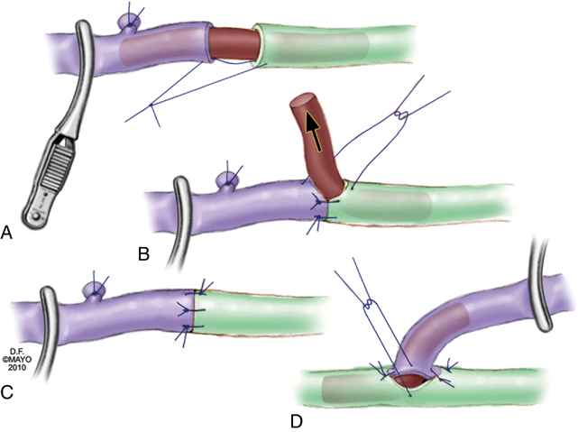
Microsurgical lymphovenous anastomoses in the leg are performed between lymph vessels of the superficial medial lymphatic bundle and usually with superficial veins. The most commonly performed techniques are direct end-to-end, end-to-side, or side-to-end microsurgical anastomoses using high-power magnification, supermicroscopic techniques, and fine 9–0 to 11–0 monofilament sutures. Important technical improvements include using 6–0 or 7–0 monofilament sutures to stent the lymph vessel or a small vein for easier and better anastomosis (Figures 1 and 2). The other is the side-to-end lymphovenous anastomosis technique that, in theory, can keep the lymph vessel patent even if the vein occludes after surgery. This might prevent progression of the lymphedema as a complication of surgical treatment.


Stay updated, free articles. Join our Telegram channel

Aug 25, 2016 | Posted by in CARDIOLOGY | Comments Off on Surgical Treatment of Lymphedema

Full access? Get Clinical Tree

Get Clinical Tree app for offline access