Surgical Interventions for Congenital Heart Disease

53 Surgical Interventions for Congenital Heart Disease


Our understanding of the complexities of congenital heart disease, a deviation from normal cardiac anatomic development that affects 8 in 1000 births, has progressed immensely since the establishment of the Board of Pediatric Cardiology in 1961. Improvements in diagnostic imaging (including echocardiography, cardiac angiography, and magnetic resonance imaging) and innovations in surgical repair techniques have resulted in greatly improved outcomes for children with congenital heart disease. This chapter provides a broad overview of the most common congenital heart lesions and the role of surgical interventions.


Embryologic development of the heart begins with the fusion of angiogenic cell clusters within the splanchnic mesoderm layer of the primitive embryo to form the heart tube at 18 to 21 days of gestation. The heart begins to rhythmically contract as early as day 17, once functional units of the myocytes begin to form. Myocardial growth proceeds with segmentation and looping of the heart tube and cellular differentiation and migration along the embryologic axes, with the establishment of laterality, and with the organization of the primitive cells into a sophisticated organ. Deviations from this complex process of cardiac development lead to congenital cardiac anomalies, with clinical presentations that in some cases occur in the immediate postnatal period and in other cases, young adulthood.


Therapy for congenital heart disease has evolved with surgical and nonsurgical innovations. The development of pediatric cardiac surgery has led to the survival of many children with complex congenital heart disease. These successes have depended on improved diagnoses, advances in surgical technique, and the development of a means for extracorporeal circulation–cardiopulmonary bypass (CPB). Complex repairs for previously fatal lesions such as transposition of the great arteries (TGA) and hypoplastic left-sided heart syndrome (HLHS) have become routine, with declining mortality rates and improved long-term outcomes. The development of transcatheter procedures has made therapeutic cardiac catheterization a viable alternative to surgery for specific congenital cardiac lesions (see Chapter 52).


Surgical Treatment


Many congenital heart lesions can be surgically repaired, meaning that approximately normal anatomy and physiology are established. The closure of atrial septal defects (ASDs) and ventricular septal defects (VSDs) can, when successfully accomplished in a young patient, eliminate the long-term consequences of the defect. Other lesions, most notably the single-ventricle defects, cannot be repaired but can be successfully palliated with operative modifications that provide a viable alternative cardiopulmonary physiology. These palliative operations can also be useful as a bridge to complete repair after a period of growth and development.


Palliative Surgical Procedures


The Blalock-Taussig shunt was first performed in 1944 at the Johns Hopkins Hospital to supply blood to the branch pulmonary arteries of a severely cyanotic patient with tetralogy of Fallot. The shunt was created by dividing the right subclavian artery and directly anastomosing it to the right pulmonary artery. Since that time, the creation of a systemic-to-pulmonary artery shunt (a “BT shunt”) has been used for a variety of congenital heart defects with severe pulmonary artery stenosis or atresia as a bridge to more definitive surgical correction. The technique of creating a BT shunt has been modified significantly by the use of an interposition graft, most commonly made of expanded polytetrafluoroethylene (ePTFE; Gore-Tex), between the systemic and pulmonary vessels. The “modified BT shunt” operation can be performed through a lateral thoracotomy or a median sternotomy and can be performed on either the right or left. A significant development in congenital heart surgery has been the increasing trend toward definitive repair at an earlier age, including the neonatal period. For this reason, the use of BT shunts is decreasing, but they still remain an important tool for the palliation of cyanotic heart lesions.


The pulmonary artery (PA) band is an important palliative procedure for congenital heart lesions in which there is excessive pulmonary blood flow. The PA band is simply a Teflon tape looped around the main pulmonary trunk and tightened to restrict pulmonary blood flow. The PA band was originally developed to treat large VSDs by decreasing the left-to-right shunt. Currently, the PA band is used most commonly for single-ventricle lesions with nonrestrictive pulmonary blood flow. The band is used to balance the systemic and pulmonary circulations and to protect the pulmonary vasculature from prolonged exposure to high pressure, which could lead to a fixed increase in pulmonary vascular resistance and irreversible pulmonary hypertension. The PA band in these patients with single ventricles is a bridge to eventual “Fontan physiology,” or total cavopulmonary blood flow. There are still some instances where a PA band is useful for the treatment of a VSD, most commonly in patients with multiple VSDs, also known as a “Swiss cheese septum.”


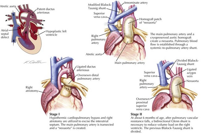
The superior bidirectional cavopulmonary anastomosis, often referred to as the Glenn shunt, is the second stage in the palliation of all or most defects of the single-ventricle type. This shunt involves the disconnection of the superior vena cava (SVC) from the right atrium and a direct anastomosis of the SVC to the right pulmonary artery (Fig. 53-1). This allows all of the systemic venous return from the upper body to flow directly to the lungs. The Glenn shunt is typically performed between 4 and 9 months of age, allowing for enough lung maturity to permit this passive blood flow. Patients will not be fully saturated following the Glenn shunt, since the venous return from the lower body still returns to the heart to mix with the fully oxygenated pulmonary venous blood. Depending on the particular cardiac defect, the procedure may be combined with the removal of a BT shunt or removal of a PA band with division of the main pulmonary trunk from the heart. In either instance, the attendant decrease in the volume load on the heart is beneficial to the function of the single ventricle and its long-term durability.


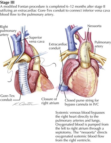
The ability of the pulmonary circulation to accept the entire cardiac output passively is limited and thus the rationale for creating total cavopulmonary circulation in two stages. The Glenn is performed in the first year of life, and the completion of the Fontan is done in the second or third year of life. There are two distinct techniques for completing the Fontan, the lateral tunnel and the extracardiac conduit. The lateral tunnel involves creating a tunnel inside of the right atrium that extends from the opening of the inferior vena cava (IVC) to the opening of the SVC, which is in turn connected to the right pulmonary artery, thus baffling all of the systemic venous return to the lungs. The extracardiac Fontan is created by disconnecting the IVC from the right atrium and sewing a graft of ePTFE from the open end of the IVC to the pulmonary arteries (Fig. 53-2). Using either strategy, a direct connection of the IVC flow to the lungs establishes total cavopulmonary circulation, with the result being that after repair, the patient should have nearly normal oxygen saturation.


Jun 12, 2016 | Posted by in CARDIOLOGY | Comments Off on Surgical Interventions for Congenital Heart Disease

Full access? Get Clinical Tree

Get Clinical Tree app for offline access