Aortic Stenosis

34 Aortic Stenosis


The outflow of blood from the left ventricle can become limited because of abnormalities of the aortic valve or narrowing of the aortic outflow tract either below or above the aortic valve. The leaflets of the aortic valve form three pocket-like cusps of approximately equal size that separate the left ventricle from the aorta. The normal aortic valve opens completely during systole, allowing unimpaired ejection of blood from the left ventricle. Closure of the aortic valve prevents retrograde blood flow from the aorta into the left ventricle and allows the left ventricle to fill solely from the left atrium in preparation for the next cardiac cycle. The most common cause of aortic outflow obstruction is valvular aortic stenosis; that is, an abnormality within the valve apparatus that obstructs flow by impairing valve mobility and opening.


Nonvalvular obstruction of left ventricular (LV) outflow usually results from a congenital abnormality, such as a membrane, that may exist above or below the valve. Hypertrophic obstructive cardiomyopathy (HOCM), formerly called idiopathic hypertrophic subaortic stenosis, produces a dynamic subaortic obstruction and is the focus of Chapter 19.


Etiology and Pathogenesis


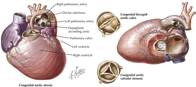
The etiology of valvular aortic stenosis varies with the patient’s age at presentation. In childhood, valvular congenital abnormalities are the usual cause. The aortic valve may be unicuspid, bicuspid, tricuspid, or, rarely, even quadricuspid (Fig. 34-1). Unicuspid valves usually are severely narrowed at birth and produce symptoms in infancy. Bicuspid and malformed tricuspid valves rarely cause symptoms during childhood. More frequently, the abnormal architecture of bicuspid and malformed tricuspid valves alters flow patterns across the valve, slowly traumatizing the leaflets, leading to progressive fibrosis, calcification, and stenosis between ages 40 and 70 years.


Acquired abnormalities from senile, calcific degeneration of a previously normal valve predominate in patients diagnosed after age 70 years, with a prevalence of 3% to 5% in patients over 75 years old (Fig. 34-2). The pathophysiology of degenerative, calcific aortic stenosis is an area of ongoing investigation. Although it shares some features and risk factors with atherosclerosis (accumulation of atherogenic lipoproteins, evidence of low-density lipoprotein oxidation, inflammation, and microscopic calcification), there are important differences. These include the presence of osteochondrogenic differentiation markers on the surface of valvular endothelial cells, essentially leading to “bone formation” in the valve, as well as a correlation between aortic stenosis and low serum levels of fetuin-A, a serum-based inhibitor of calcification.


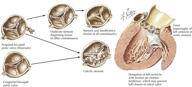
Rheumatic involvement of the aortic valve, less prevalent today in the United States than a generation ago, typically results in a combination of stenosis and regurgitation, usually with concomitant mitral valve disease. The rheumatic valve is characterized by commissural fusion and calcification, whereas the more common degenerative aortic stenosis shows calcification progressing from the base of the cusps toward the leaflets, generally sparing the commissures (see Fig. 34-2). Rheumatic aortic stenosis generally presents between ages 30 and 50. Less common causes of aortic stenosis include obstructive vegetations from endocarditis, history of radiation therapy, and rheumatoid involvement with severe nodular thickening of the valve leaflets. Aortic stenosis may also be associated with systemic diseases including Paget’s disease, Fabry’s disease, ochronosis, and end-stage renal disease.


Bicuspid aortic valve disease merits special consideration given its prevalence (approximately 1% in the general population) and the frequent association of bicuspid aortic valve disease with genetic disorders that lead to enhanced elastolysis of the aortic wall and accelerated apoptosis of smooth muscle cells of the aortic media. This abnormality leads to a reduced aortic elasticity and dilatation of the annulus, aortic root, and ascending aorta with an increased risk of aortic dissection. Because of this defect at the arterial wall level, many experienced centers now approach patients with bicuspid aortic valve and aortic dilatation similarly to patients with Marfan’s syndrome, considering elective repair when the aortic diameter approaches 4.5 to 5.0 cm, instead of the traditional threshold size of 5.5 cm.


Clinical Presentation


Valvular aortic stenosis is often asymptomatic for years. Progressive and ultimately severe pressure overload imposed by the valve stenosis results in the development of concentric LV hypertrophy (LVH). This compensatory adaptation lowers wall stress and maintains forward flow but also leads to detrimental effects, including an abnormal diastolic filling pattern and subendocardial ischemia.


Classic symptoms of aortic stenosis are angina, syncope, and dyspnea, the latter being a manifestation of congestive heart failure (CHF). The average survival without valve replacement is 5 and 3 years in patients who present with angina or syncope, respectively. The most concerning presentation is CHF, because those patients have an average survival without valve replacement of less than 2 years.


Angina occurs in two thirds of patients with severe aortic stenosis, and approximately half of these have significant coronary obstructions. In the absence of important coronary obstructions, angina is caused by subendocardial ischemia induced by increased wall thickness with a relatively decreased capillary density, prolonged ejection time, and increased LV end-diastolic pressure, which reduces the diastolic transmyocardial perfusion gradient.


Syncope in patients with important aortic stenosis typically occurs during physical exertion. Exertion reduces systemic vascular resistance, while the necessary increase in cardiac output is blunted by the fixed valve obstruction. This combination results in cerebral and cardiac hypoperfusion. Moreover, the greatly increased LV systolic pressure during exertion activates ventricular baroreceptors that may cause an exaggerated vasodepressor response, further reducing cerebral perfusion. Common arrhythmias such as atrial fibrillation or atrioventricular conduction abnormalities may reduce LV filling and cardiac output, resulting in syncope. Life-threatening arrhythmias, such as ventricular tachycardia or fibrillation, though uncommon, may occur in patients with aortic stenosis and result in syncope occurring at rest or with exertion and the potential for sudden cardiac death.


CHF can develop because of diastolic dysfunction, related to severe LVH with delayed ventricular relaxation and decreased compliance. Systolic dysfunction with progressive ventricular dilatation may occur late in the disease course. To compensate for the LV pressure load, the left atrium hypertrophies and develops vigorous contractions that allow adequate LV filling despite increased LV end-diastolic pressure. However, as the disease progresses or with physical activity, left atrial pressure increases further, leading to higher pulmonary venous pressures and eventually to pulmonary congestion and edema. Pulmonary edema may develop abruptly during activity or with the loss of atrial function, as in atrial fibrillation.


Aortic stenosis may be associated with gastrointestinal bleeding from angiodysplasia (so called Heyde’s syndrome, which is associated with a mild form of acquired von Willebrand’s disease), or embolic events from detachment of small calcium deposits. Infective endocarditis is also a risk in patients with aortic stenosis and can have many morbid complications.


Physical Examination


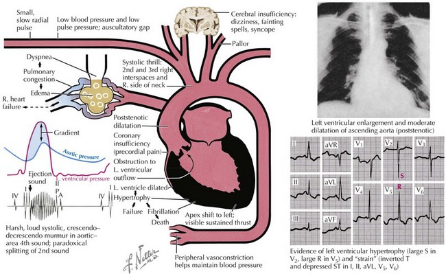
Although noninvasive imaging provides an excellent tool for evaluation of patients with aortic stenosis, this condition can be assessed by a careful physical examination. One of the most notable findings in severe aortic stenosis is a decreased pulsation of the carotid arteries and a slowed arterial upstroke (pulsus parvus et tardus), with the maximum carotid upstroke noticeably delayed after the apical impulse (Fig. 34-3). A marked vibration or shudder may also be felt in the carotid artery. The jugular venous pressure is not elevated unless CHF is present. In mild aortic stenosis the jugular venous pulsations may be unremarkable, whereas late in the disease a prominent “v” wave may occur from tricuspid insufficiency caused by pulmonary hypertension and bulging of the hypertrophied septum into the right ventricle. As the degree of valve stenosis progresses, the LV apical impulse becomes displaced inferiorly and laterally, with a palpable presystolic pulsation (palpable S4). If the apical impulse is hyperdynamic, concomitant aortic or mitral insufficiency should be considered.


The first heart sound is usually normal, but the second heart sound may have a reduced aortic closure sound or be single because of the absence of the aortic component from immobile aortic leaflets; it may also be paradoxically split from a marked delay of LV ejection. There is often a fourth heart sound (S4), reflecting the reduced LV compliance during atrial contraction, and a third heart sound (S3

Stay updated, free articles. Join our Telegram channel

Jun 12, 2016 | Posted by in CARDIOLOGY | Comments Off on Aortic Stenosis

Full access? Get Clinical Tree

Get Clinical Tree app for offline access