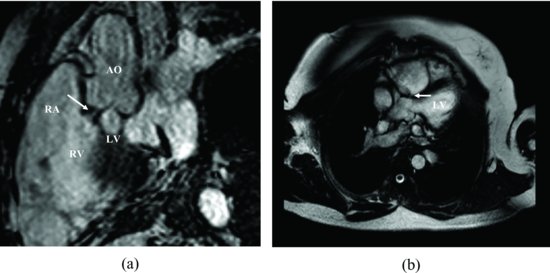
Figure 34.2 A small shunt can be seen in the magnetic resonance images (arrows point) (a and b). AO, aorta; LV, left ventricle; RA, right atrium; RV, right ventricle.
The patient’s shunt is hemodynamically insignificant and is unlikely to cause symptoms or progress.
Discussion
There are various types of VSD; the infundibular ventriculoseptal-type occurs less frequently representing 5–7% of VSDs [1]. This type of septal defect is located under both semilunar valves, at the fibrous continuity between the pulmonary and aortic rings, with the infundibular septum either partially or completely missing.
Stay updated, free articles. Join our Telegram channel

Full access? Get Clinical Tree


