Figure 46.1
The patient is intubated with a double-lumen tube and placed in a lateral decubitus position. The sterile field is prepared to include the upper arm and scapula, with skin preparation reaching from the base of the skull down to the iliac crest and past the spinal processes posteriorly and the midline anteriorly, encompassing the neck, manubrial notch, and sternum. The lower arm should be draped to just above the elbow to permit a gentle pull of the upper arm by the second assistant to elevate the scapula, if necessary. The surgeon stands at the patient’s back, the first assistant across the table, and the second assistant cephalad to the surgeon. The patient’s arm is placed on a sterilely draped padded arm rest. A long posterior thoracotomy is made starting midway between the spinal processes and the posterior aspect of the scapula, and is extended forward in a gentle arch 2–3 cm below the inferior angle of the scapula until the anterior border of the latissimus dorsi muscle is reached. As a modification, according to Tatsamura, this incision may be continued upward to above the nipple level to access more anteriorly located structures and to permit adequate mediastinal lymphadenectomy

Figure 46.2
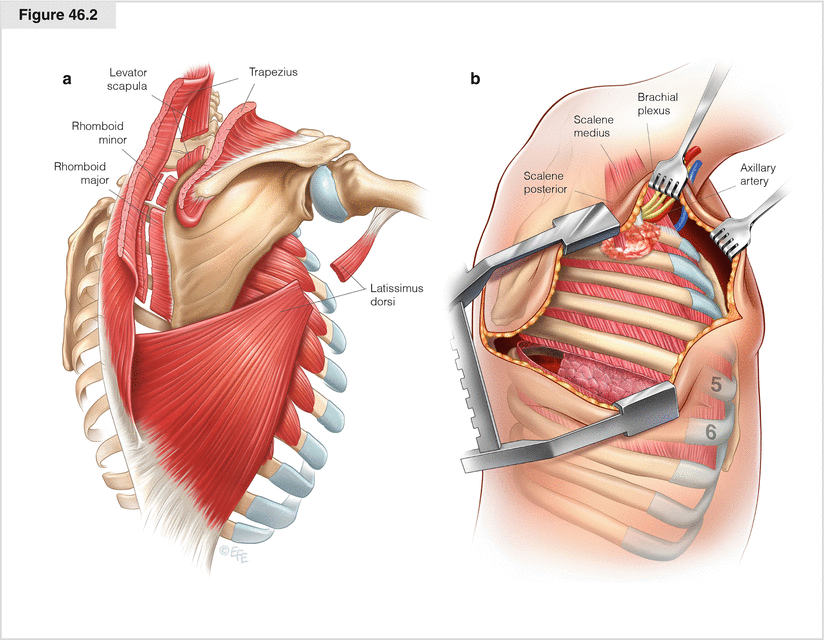
Anteriorly, the latissimus dorsi and the fascia posterior to the anterior serratus muscle are incised along their posterior edges (a). Posteriorly, the trapezius muscle is divided along the full length of the incision, then the lesser and greater rhomboid muscles and the levator muscle of the scapula. Care should be taken not to injure the dorsal scapular nerve and satellite scapular artery at this point. The anterior serratus muscle fibers along the fifth intercostal space are divided, and the pleural cavity is entered after the intercostal muscles are divided (b). This approach allows the assessment of resectability and better visualization of the upper lobe. Next, a retractor is placed to displace the scapula upward and forward and the sixth rib downward. At this point, the brachial plexus, axillary artery, and middle and posterior scalene muscles are visible

Figure 46.3
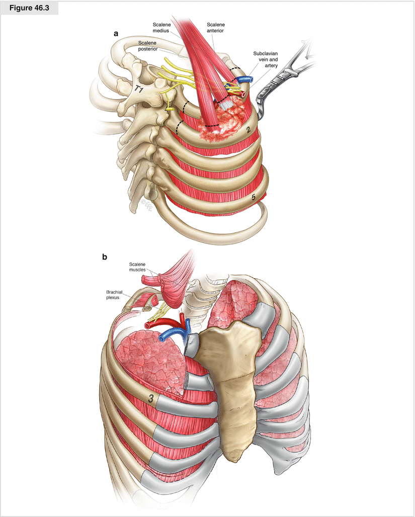
(a, b) Simultaneous bimanual palpation at this point helps determine the extent of tumor infiltration to the chest wall and into the thoracic inlet. As shown here, the first rib and the cranial portion of the second are infiltrated. It is important that negative resection margins are obtained. Therefore, the intercostal muscles of the second intercostal space are divided close to the third rib or the third rib is included in the resection. With a Brunner rib shear, the second rib is divided first anteriorly and then posteriorly. Intervening intercostal neurovascular pedicles are suture ligated or clipped and then divided. The middle and posterior scalene muscles are transected with cautery, providing access to the posterior angle of the first rib. After the angle of the invaded first rib is dissected to obtain tumor-free margins, the rib is transected with a right angle rib shear. If no further infiltration is present in the brachial plexus, subclavian artery, and vein, the anterior scalene muscle is transected and the first rib divided with a 60°-angle rib shear. When dividing the anterior scalene muscle, care must be taken not to sever the subclavian vein, which runs ventrally to this muscle. This may be avoided by the operator placing a finger between the muscle and vein and transecting the muscle directly at the point of its insertion onto the first rib. The rib should be cut to the level of the costocartilage anteriorly. Following resection including the first and second rib, the segment of chest wall is dropped into the chest cavity and the procedure is completed by lobectomy and mediastinal lymphadenectomy

Figure 46.4


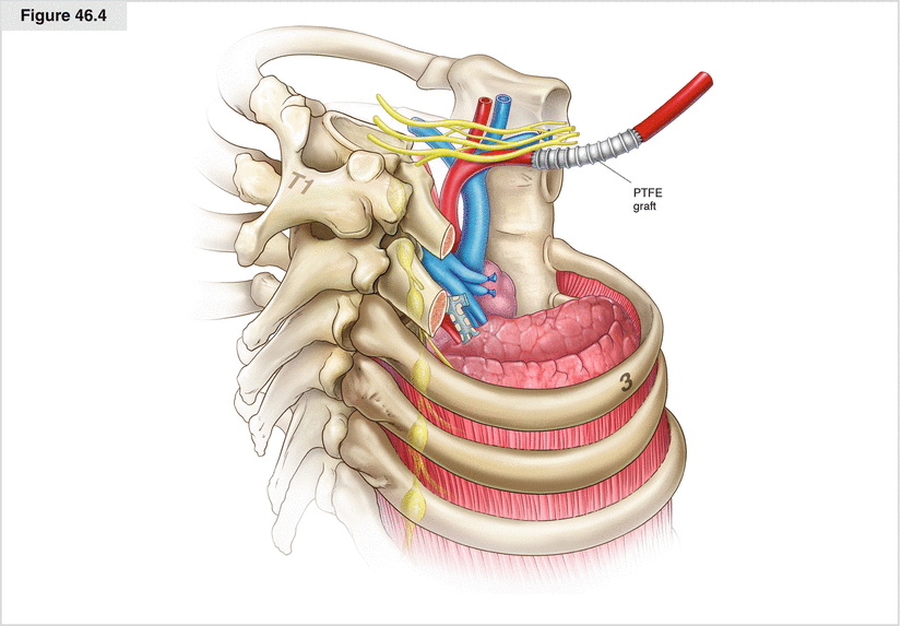
If tumor invades the most posterior aspect of the first and second ribs near the transverse ligaments, as may be confirmed intraoperatively by a frozen section of uncalcified periosteum, the transverse process along with the lateral cortex of the vertebrae may be transected with an osteotome. Up to one quarter of the vertebral body may be removed without affecting stability. The sympathetic chain is then divided above and below the tumor mass, and the stellate ganglion is resected. Most commonly, tumor invasion of the brachial plexus is limited to the first thoracic and eighth cervical nerve roots. In these cases, the lower trunk of the brachial plexus should be divided. To prevent cerebrospinal fluid leakage, the nerve roots should be secured with a ligature before being transected at the intervertebral foramen. If the subclavian vein and artery are infiltrated by tumor, special operative measures are indicated. The vein may simply be sutured and ligated; the artery should be cross-clamped. Following adequate systemic heparinization, revascularization by end-to-end anastomosis or interposition of a ring-fortified polytetrafluoroethylene (PTFE) graft (6 or 8 mm) should be performed, as shown here. However, such procedures may be very hazardous if attempted through a posterior approach; as a safer alternative, such maneuvers should be performed through an additional anterior approach (see Fig. 46.5)
Stay updated, free articles. Join our Telegram channel

Full access? Get Clinical Tree


