Sudden Cardiac Death in Athletes

69 Sudden Cardiac Death in Athletes


More Americans than ever engage in athletics—from occasional exercisers to highly competitive, high-profile amateur and professional athletes. Sudden death in athletes, especially from cardiovascular causes, is an uncommon occurrence, with an estimated prevalence of 1 in 200,000 athletes per year. Despite this low prevalence, sudden death in athletes is a noteworthy event for the media and the community for many reasons: athletes often enjoy celebrity status in American culture, athletes are typically young, and their deaths are deemed to be tragically premature. Moreover, their deaths contradict popular perception that young athletes personify health and vitality. As the number of highly trained athletes increases, more attention has been placed on the cardiovascular assessment of athletes as well as physicians’ abilities both to diagnose potentially lethal diseases and to prevent early death.


Causes of Sudden Cardiac Death in Athletes


Epidemiologic evidence suggests that sudden death in young athletes, ages 12 to 40 years, occurs more often in males than females, and that participation in basketball and football is implicated in greater than two thirds of cases. Of deaths not related to trauma or accidents, 90% occur during or immediately following a training workout or an athletic event. While the most common cause of death in athletes at least 35 years of age is coronary atherosclerotic disease, the two most common causes of cardiovascular death in younger athletes are hypertrophic cardiomyopathy (HCM; 36%) and anomalous origin of coronary arteries (17%). Among other cardiovascular causes of death, rupture of an aortic aneurysm associated with Marfan’s syndrome, mitral valve disease, dilated cardiomyopathy, aortic stenosis, and arrhythmogenic right ventricular cardiomyopathy account for 2% to 6% of cases, while drug abuse, long QT syndrome, cardiac sarcoidosis, and other cardiovascular causes account for 0.5% to 1% of cases. Commotio cordis represents the most frequent cause of traumatic death in athletes and is the etiology of death in approximately 20% of cases. Other causes of traumatic death include head and spine injuries from bodily contact, and even vascular injury to coronary, vertebral, and internal carotid arteries from incoming projectile objects such as balls and hockey pucks. The challenge for physicians evaluating athletes for cardiac risk is to rule out potentially lethal pathology that could lead to sudden cardiac death. This chapter focuses on four of the common causes of sudden cardiac death that must be considered in evaluation of athletes.


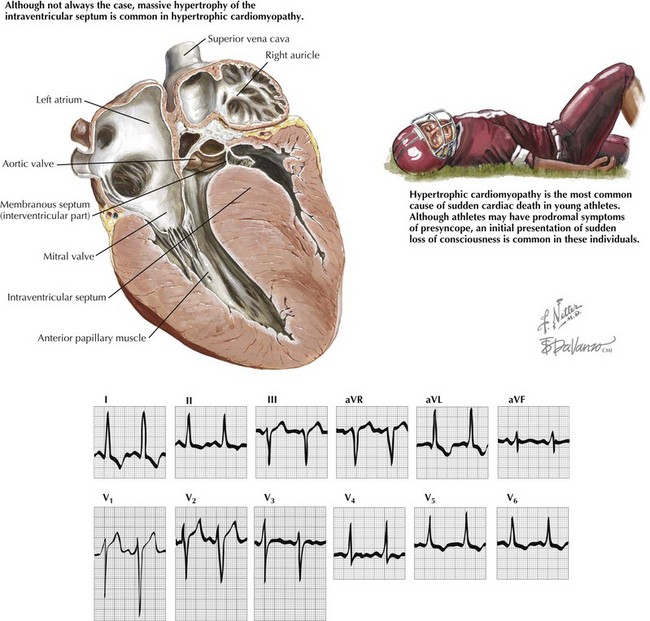
Hypertrophic Cardiomyopathy


HCM, transmitted in an autosomal-dominant fashion, results from mutations in any of 10 genes that encode specific constituents of the cardiac sarcomere. Over 200 mutations have now been identified. The most common mutations involve the β-myosin heavy chain and myosin-binding protein C. The estimated prevalence of HCM in the healthy, young, general population has been estimated at 0.17% in the United States. Worldwide, the prevalence of HCM in athletes is probably lower, estimated at approximately 0.07%. Although the prevalence is low, early diagnosis of HCM is crucial for those engaged in competitive sports because of the risk of sudden cardiac death. The risk of sudden cardiac death in individuals with HCM varies considerably, depending on the causative mutation and other genetic and environmental factors that are not fully understood. Because it is not possible at present to define HCM individuals who have minimal risk, and because competitive and even strenuous exercise are associated with sudden cardiac death across the spectrum of HCM, most experts recommend against competitive athletics in individuals with HCM. Furthermore, individuals with HCM identified through this kind of screening should also be considered for potentially lifesaving therapy, including cardioverter-defibrillator implantation, depending on numerous factors.


Ventricular tachycardia and ventricular fibrillation are the most frequent etiologies of sudden cardiac death in patients with HCM. The six features associated with greatest risk for sudden cardiac death are prior cardiac arrest or sustained ventricular tachycardia, family history of one or more premature HCM-related deaths, syncope, hypotensive blood pressure response to exercise, multiple or prolonged episodes of nonsustained ventricular tachycardia on ambulatory monitoring, and left ventricular (LV) wall thickening greater than 30 mm (Fig. 69-1).
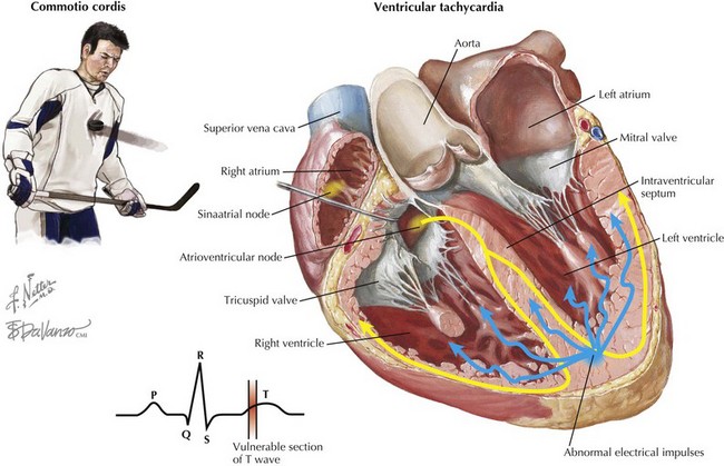


Because the hearts of highly conditioned athletes are often enlarged and proportionally hypertrophied, it is equally important not to label individuals with an “athlete’s heart” as having HCM. Cardiac enlargement and hypertrophy in athletes may simply represent a physiologic response to increased myocardial demand in training, and after cessation of vigorous training these changes can resolve over time. Doppler echocardiography can help distinguish normal athletes’ hearts from HCM. Normal diastolic filling patterns are generally present in enlarged and hypertrophied athletes’ hearts, whereas the hearts of patients with HCM show abnormal diastolic function including decreased early peak flow velocity, slowed deceleration of early diastolic flow velocity, and increased late peak flow velocity associated with atrial systole. Besides echocardiography, metabolic exercise stress testing can distinguish HCM from athlete’s heart; specifically, athletes with LV hypertrophy without HCM can achieve a peak maximum oxygen consumption of 50 mL/kg/min, but athletes with true HCM generally cannot. Cardiac MRI has also emerged as a useful tool in HCM diagnosis and may be even more sensitive than echocardiography in identifying areas of hypertrophy, especially in the anterolateral wall.


Traditionally, screening tools for HCM include a history and physical examination along with ECG and echocardiogram. Screening for HCM typically begins at age 12, unless there are factors that would provoke an earlier or a more in-depth evaluation, including a family history of premature death related to HCM, onset of symptoms, other evidence of early LV hypertrophy, or participation in competitive athletics requiring intense physical training. Generally, screening with traditional methods has been recommended every 12 to 18 months between ages 12 and 21, when adult physical maturity is achieved. Evidence indicates that phenotypic appearance of HCM can occur well into adulthood and, in some mutations, as late as in the fifth or sixth decade of life. Thus, the absence of morphologic characteristics of HCM at early adulthood should not be viewed as conclusively ruling out HCM for either patients or practitioners. Recent recommendations have suggested continuing screening with serial ECGs and echocardiograms at 5-year intervals into midlife and perhaps beyond.


Because patients with HCM may not manifest symptoms or signs of hypertrophy until later in life and because phenotypic expression is so variable even among family members, athletes with a positive family history for HCM should be considered for further testing, to include echocardiography and MRI. Genetic analysis of families with certain HCM mutations has demonstrated that not all individuals who carry the mutation show phenotypic or imaging evidence of HCM. Thus, many experts now also recommend genetic testing for individuals with a family history of HCM. Although some genotype-phenotype correlation studies suggest that mutations in some genes are “malignant” (associated with a high incidence of sudden cardiac death) while other mutations are “benign” (associated with a normal life expectancy), other studies have illustrated that patients with either the malignant or benign genotype can have a variable clinical course. Because of this variability, athletes identified as having any mutation associated with HCM should be considered high risk for sudden death.


Coronary Artery Anomalies


Congenital coronary artery anomalies account for a significant proportion of sudden death in athletes in the United States, especially in athletes age 35 or younger. Screening for these abnormalities is difficult, because initial clinical suspicion is lacking and routine testing is unable to identify this particular abnormality. Individuals with anomalous coronary arteries often have repetitive episodes of myocardial ischemia and/or microinfarcts that can result in an increased risk for ventricular arrhythmias. Ventricular tachycardia and ventricular fibrillation are the most common causes of sudden cardiac death in individuals with congenital coronary anomalies. Other potential causes of death include obstruction or closure of a slitlike ostium, spasm of the anomalous coronary artery, compression of the anomalous artery (due to vigorous myocardial contraction), and endothelial injury. Early identification of coronary abnormalities is particularly important, because intense physical activity should be avoided before surgical correction. At this time, coronary artery bypass grafting remains the therapy of choice, but other investigations are exploring the efficacy of reimplanting the anomalous vessels into the proper coronary sinus.


A review of two large U.S. and Italian registries demonstrated that the most common coronary artery anomalies included abnormal origin of the left main coronary artery from the right aortic sinus and origin of the right coronary artery from the left sinus.


On pathologic examination, hearts with this anomaly demonstrate a sharp takeoff of the artery at the ostium of the improper aortic sinus as well as an anatomic course passing between the aorta and the pulmonary trunk. In some specimens, the proximal portion of the anomalous artery was intramural and contained within the aortic wall. Many specimens had evidence of acute ischemia, including contraction band necrosis, wavy fibers, and early neutrophilic infiltrate in the myocardial territory supplied by the anomalous artery. There was also evidence of chronic ischemic injury and patchy replacement-type fibrosis. These pathologic specimens support the hypothesis that both acute and chronic ischemic injuries predispose athletes with anomalous coronary arteries to fatal ventricular arrhythmias. The majority of patients who died as a result of having anomalous coronary arteries were male and 60% were Caucasian, with the others being African American or Asian. Deaths have occurred at all levels of competitive athletics, from teenagers in amateur recreational sports to collegiate and professional athletes.


Although their diagnosis remained undetected, more often than not the athletes admitted to prior signs and symptoms of cardiovascular disease, including syncope, chest pain, dizziness, and palpitations. Of the 27 athletes identified in one review as having died from an anomalous origin of a coronary artery, 4 had reported at least one prior episode of syncope, and in 2 athletes, the syncopal episode occurred within 11 to 24 months of death. Recurrent syncope had occurred in 2 individuals who later died. Five athletes experienced chest pain, usually during physical exertion. In some cases, the chest pain occurred within a few days of death, while in others it occurred within 24 months of death. The presence of symptoms should alert the clinician to perform further evaluation. Importantly, routine noninvasive examinations may be misleading; 9 patients had ECGs performed before death, all of which were within normal limits. Six of the athletes underwent exercise stress testing, all the results of which were within normal limits. Two athletes had two-dimensional echocardiograms performed that were both normal. CT angiography is now capable of defining anomalous coronary arteries and should be considered in individuals with worrisome symptoms but normal examinations, ECGs, echocardiograms, and stress tests.


Commotio Cordis


Commotio cordis refers to a blunt, nonpenetrating blow to the chest wall that results in sudden cardiac death. Based on experimental data, it is thought that commotio cordis primarily occurs only when chest trauma occurs just before the peak of the T wave during repolarization. In a swine model of commotio cordis, it was demonstrated that the vulnerable point in the cardiac cycle was between 15 and 30 ms before the peak of the T wave. In this model, 90% of the chest blows (either with a ball or a wooden bat) during this portion of the cardiac cycle induced ventricular fibrillation. Blows to the chest outside of this time period did not induce ventricular fibrillation but did produce brief episodes of polymorphic ventricular tachycardia. In settings of electrolyte abnormalities or underlying cardiac disease, the induction of polymorphic ventricular tachycardia may also result in ventricular fibrillation and sudden cardiac death. Complete heart block, ST-segment elevation, and left bundle branch block were observed when impacts to the chest occurred during the QRS complex and not during the vulnerable phase. Complete heart block was only observed with chest blows during the QRS complex and not with impacts at other times during the cardiac cycle. Coronary angiography performed after blunt injury did not reveal significant coronary artery abnormalities, such as spasm, dissection, or stenosis, consistent with the idea that the cause of sudden cardiac death was arrhythmic (Fig. 69-2).


Commotio cordis has been reported in athletes engaged in sports involving a projectile ball, including baseball, softball, cricket, basketball, soccer, and lacrosse, as well as other sports such as hockey and martial arts. In a large series of 128 documented cases of commotio cordis, 58% of events occurred during baseball or softball games whereas 16% of events occurred during hockey games. Most cases of commotio cordis during athletic events involved a projectile causing blunt force to the chest wall. Most of these projectiles were balls composed of a solid core rather than an air-filled ball (i.e., soccer or basketball). Nonprojectile causes of commotio cordis included bodily contact between players with a shoulder, elbow, knee, or foot. Of particular importance is that a large percentage of commotio cordis events occurred during recreational activities outside of organized sports, including those residential backyards and homes. One such episode included a 5-year-old child who died after being struck in the chest by a plastic sledding saucer; another involved a man who died after his friend struck him on the chest to help alleviate his hiccups.


Survival of commotio cordis is probably related to timing of resuscitative efforts. Of the 128 cases documented in this series, 106 had cardiopulmonary resuscitation performed, mostly by trained professionals, including physicians, nurses, firefighters, and emergency medical services technicians. Of the 68 cases in which cardiopulmonary resuscitation was initiated within approximately 3 minutes or less, 17 patients survived. When cardiopulmonary resuscitation was initiated after 3 minutes, only 1 person out of 38 survived.


Although commotio cordis can occur with both hard and soft objects, there does seem to be a correlation between the object’s hardness and the induction of ventricular fibrillation. In the swine model discussed previously, a wooden object simulating a bat and four types of baseballs with varying degrees of hardness were used to strike the chest wall: very soft (designed for 5- to 7-year-olds), medium-soft (designed for 8- to 10-year-olds), least soft (designed for children 11 years and older), and regulation Little League. There was a significant difference between the very soft baseball and the regulation Little League baseball, with the very soft baseball inducing ventricular fibrillation in 2 of 26 blows to the chest and the regulation baseball inducing ventricular fibrillation in 8 of 23 impacts. There was also a significant difference in induction of ventricular fibrillation between the wooden object and all baseballs. Blows to the chest with the wooden object resulted in ventricular fibrillation 90% of the time, whereas ventricular fibrillation occurred after impact with baseballs at a maximum frequency of 35% with the regulation baseballs. Unfortunately, commercially available protective equipment does not provide a total safeguard against death. The impact from a baseball traveling at 40 miles per hour can induce ventricular fibrillation up to 49% of the time despite the use of commercially available protective chest gear. Similarly, lacrosse chest protectors allowed ventricular fibrillation to occur up to 50% of the time when impact was delivered with a lacrosse ball traveling at 40 miles per hour.


Jun 12, 2016 | Posted by in CARDIOLOGY | Comments Off on Sudden Cardiac Death in Athletes

Full access? Get Clinical Tree

Get Clinical Tree app for offline access