Subintimal Recanalization for Lower Limb Occlusive Disease


Subintimal Recanalization for Lower Limb Occlusive Disease


Andrew L. Tambyraja and Amman Bolia


Subintimal recanalization was originally described in 1989 as a novel technique to treat chronic total occlusions of the femoropopliteal arterial segment. The need for an alternative angioplasty strategy arose from patients with anatomically complex, long, heavily calcified lesions in whom traditional primary endoluminal angioplasty was often met with difficulty and technical futility.


Subintimal recanalization offers an effective alternative endovascular solution for such chronic arterial occlusive disease, particularly in those with critical limb ischemia who are poor candidates for surgical bypass. The technique challenges established principles of conventional endoluminal arterial angioplasty by advocating a deliberate catheter-initiated dissection into a neoanatomic plane between the arterial intima and media. This dissection is propagated along the length of the occlusion and then directed back into the arterial true lumen beyond the occlusion. The subintimal plane is then balloon dilated to enlarge into the preferred channel for blood to travel through. Concerns that the exposed media in the subintimal channel creates a potent thrombogenic surface have not been borne out in studies of long-term patency.


The technique has been slow to gain general acceptance, and it continues to have its skeptics, yet its application has now been extended to the iliac and crural vessels and is acknowledged as a useful tool in the treatment of critical limb ischemia. For subintimal recanalization to be successful, it is critical that alongside good inflow there must be good runoff to support fast flow through the new subintimal channel.


Subintimal recanalization is suitable for acute or subacute occlusions where the presence of soft, fresh thrombus carries a prohibitive risk of downstream thromboembolism if treated by intraluminal interventions. It is important to determine that previous attempts at subintimal angioplasty did not leave any prostheses within the vessel and do not preclude or hamper subsequent attempts at an open surgical revascularization.


Technique


An antegrade puncture of the common femoral artery is the most favorable approach to femoropopliteal lesions. However, retrograde access from the popliteal artery has also been used. Access from the contralateral femoral artery with a crossover sheath is less desirable for reasons of diminished catheter torque control and pushability, which may be further compounded in narrow aortic bifurcations and tortuous iliac anatomy. Precise antegrade puncture is especially critical in flush occlusions of the superficial femoral artery to allow adequate space for catheter and guidewire manipulation to initiate the intimal dissection. Duplex ultrasound is a very useful tool to guide puncture in these circumstances.


A 4- or 5-Fr catheter is positioned immediately adjacent to the start of the occlusion after systemic heparinization. A hydrophilic angle-tipped guidewire is carefully introduced and manipulated to form a loop as it abuts against the occlusion. With persistent probing, the loop of the guidewire creates a dissection into the subintimal space. At this point, the wire and catheter should be advanced down the potential space. The wire tends to follow a spiraling course down the vessel, and the loop might appear slightly wider than the expected diameter of the vessel. The length of wire taken up in the loop should be controlled by regularly shortening the amount of wire outside the catheter. The greater the length of wire in the loop, the greater the force being used in the dissection by the stiffer portion of the wire. However, it is critical that the loop be maintained throughout, because loss of the loop and advancement of a straight wire will fail to produce an adequate dissection, and the risk of vessel perforation or cannulation of collateral branches will be increased.


As the catheter and guidewire approach the end of the occlusion, the wire usually tends to reenter the true lumen as it encounters a normal vessel. Spinning the wire as it is advanced can facilitate reentry. This is usually heralded by a loss of resistance to the passage of the wire, which now follows the course of the true lumen. The luminal position of the catheter can be confirmed by a small injection of contrast. If the catheter remains within the dissection plane, contrast only advances a short distance in a clearly extraluminal space. In a diffusely diseased artery with extensive calcification, femoropopliteal reentry may be impossible or may be complicated by extraluminal perforation.


Perforation is reported to occur in 5% to 8% of cases and is more common than during luminal angioplasty. This risk is greatest in patients with heavily calcified vessels, very elderly patients, and current smokers. Perforation does not always signify procedural failure. In skilled hands, an alternative subintimal plane can be found more proximally and this new channel used to seal the perforation and achieve revascularization. Alternatively, the dissection can continue to propagate toward runoff vessels or major collateral branches. Although reentry into tibial vessels is feasible with good outcomes, the procedure is technically more demanding, and it may be prudent to abandon the case for a further attempt at another sitting.


An adjunct to subintimal recanalization is the advent of dedicated reentry catheters such as the Outback catheter (Cordis, Bridgewater, NJ) or the intravascular ultrasound guided Pioneer catheter (Medtronic, Minneapolis, MN). These devices incorporate a retractable curved needle at the catheter tip that can be directed and advanced at the desired point of luminal reentry to help the wire break back into the lumen.


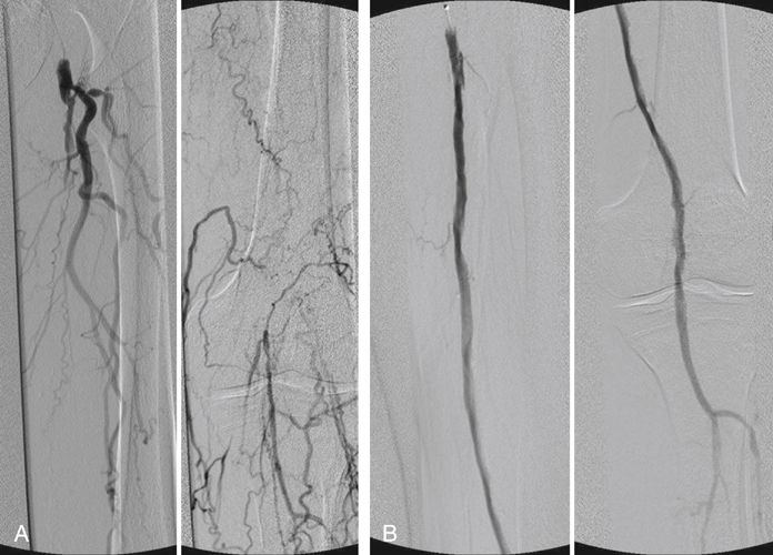
An appropriately sized balloon catheter can be used to dilate the channel with rapid segmental inflations from distal to proximal once the wire has traversed through the subintimal plane, along the occlusion, and back into the true lumen below the occlusion. After angioplasty, angiography is performed to document the characteristic appearances of a subintimal recanalization (Figures 1 and 2). Radiographic appearances demonstrate a very irregular channel. However, the most important characteristic of the angiogram is velocity of flow. Fast flow is the hallmark of a successful recanalization.


Stay updated, free articles. Join our Telegram channel

Aug 25, 2016 | Posted by in CARDIOLOGY | Comments Off on Subintimal Recanalization for Lower Limb Occlusive Disease

Full access? Get Clinical Tree

Get Clinical Tree app for offline access