Subclavian Steal Syndrome
MICHAEL J. MALINOWSKI and CHEONG J. LEE
Presentation
A 67-year-old male presented with 3 months of increasing left upper extremity numbness and tingling with strenuous use. The patient has recently also developed gangrene to the tip of his third digit within the past 3 weeks with symptoms of vertigo. According to the patient’s wife, he also has had a few episodes of unsteadiness while walking needing to support himself at points of ambulation. The patient was evaluated initially by his primary care physician and referred for evaluation. He has no evidence of prior stroke or transient ischemic attacks.
Differential Diagnosis
Although the presentation of this patient illustrates multiple symptoms pointing to subclavian artery stenosis, the differential diagnosis must be carefully reviewed. Berger’s disease in young male patients with extensive tobacco use can present with upper extremity tissue loss and ulceration, but without the proximal branch symptoms of vertebrobasilar insufficiency. When considering inflammatory etiologies, both Takayasu’s arteritis and giant cell arteritis can cause similar groupings of symptoms including upper extremity ischemia and vertebrobasilar symptomatology and should be strongly considered if there is evidence of elevated inflammatory markers, such as C-reactive protein and erythrocyte sedimentation rate, along with imaging studies featuring proximal branch vessel disease. In an older patient in the sixth or seventh decade of life, consideration must include native atherosclerotic disease especially in patients with renal failure and heavy tobacco use. Although primary Raynaud’s must be considered, the fact that disease progression has resulted in tissue loss generally rules out primary disease. The multitude of etiologies for secondary Raynaud’s disease, however, remain in the differential diagnosis.
A posterior distribution ischemic stroke must be strongly considered in this patient given the issues with coordination and balance and should be ruled out to prevent long-term morbidity of an undiagnosed cerebrovascular event. Since the patient has a subacute presentation, this scenario is unlikely embolic, although this may be considered.
In the case of subclavian stenosis, atherosclerotic disease is by far the most common etiology requiring a diameter stenosis of greater than 75%. Multivessel involvement is seen in up to 40% of patients presenting with symptomatic disease.
Workup
Full imaging of the subclavian artery by ultrasonography is limited by bony landmarks, including the clavicle. Flow dynamics, such as spectral broadening and elevated velocities, seen in the artery around the stenotic lesion should prompt further imaging, especially in concert with flow reversal in the ipsilateral vertebral artery. The gold standard of diagnostic imaging is conventional angiography provided by an arch angiogram. With the advent of advanced CT imaging and reconstruction software, computed tomography angiography (CTA) and magnetic resonance angiography (MRA) can effectively evaluate the proximal subclavian artery for stenotic lesions; however, these modalities are strictly diagnostic, as opposed to angiography that allows for assessment of translesion pressure gradients and direct endovascular therapy. Disadvantages of aortography include local arterial trauma, risk of stroke with arch manipulation, and nephrotoxicity with contrast administration. With a high percentage of these patients with proximal branch vessel disease also having concomitant coronary disease, coronary evaluation with transthoracic may be useful in assessment of cardiac function. Although the most common orientation of the transverse branch vessels involves three separate trunks of the innominate, left carotid and left subclavian arteries, aberrant arch anatomy including bovine arch orientation is present in up to 16% to 24% of patients. For evaluation of open operative revascularization, this is a less important point; however, in the context of endovascular revascularization including subclavian stenting, it is critical.
Aberrant vertebral anatomy also plays an important role not only in treatment but also in disease physiology. An aberrant left vertebral origination off the aortic arch between the left common carotid and subclavian arteries occurs in 6% of patients and an aberrant right subclavian artery origination occurs in 0.5% to 1.0% of patients.
Diagnosis and Treatment
Initial awareness of proximal subclavian stenosis may manifest itself as asymmetry in upper extremity blood pressures with a 20 mm Hg difference between upper arm brachial systolic pressure measurements. A patient presenting with vertebrobasilar insufficiency with resultant vertigo, nausea, imbalance, and diplopia, a duplex of the ipsilateral vertebral artery, will often show signs of flow reversal. Yet, this duplex finding alone is common in asymptomatic patients as well. For patients with left-sided subclavian stenosis and prior coronary bypass grafting with origination from the left internal mammary artery, subclavian-coronary steal may occur leading to myocardial ischemia, angina, and possible infarction. More proximal innominate and common carotid artery stenosis and occlusions tend to present with ipsilateral hemispheric symptoms, including aphasia and hemiparesis.
When symptoms are present a thorough physical examination, to check upper extremity pulses and associated bruits along with a comprehensive neurologic exam, should be performed followed by duplex sonography. Often angiography can be successfully used not only for diagnosis but for treatment as well. As discussed, CTA and MRA imaging is a useful adjunct to angiography, and even transesophageal echocardiography may be employed to further evaluate and characterize the level of aortic arch disease prior to intervention, but these remain diagnostic modalities only.
The main indication for treatment is the presence of symptoms, including upper extremity ischemia, vertebrobasilar symptoms, or sequelae of subclavian-coronary and subclavian-carotid steal. There are no clear guidelines of when to intervene on asymptomatic patients, if ever, but a suitable indication to consider is in a patient with high-grade left subclavian stenosis with an anticipated internal mammary artery to coronary artery bypass.
Surgical Approach
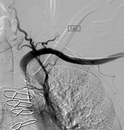
Endovascular and open techniques are both available options for treatment of symptomatic subclavian stenosis. With the advent of improved stent technology, lower profile delivery systems and knowledge of endovascular limitations including appropriate patient selection, proximal orificial subclavian stenting is considered by most as a first-line treatment. This utilizes either femoral or brachial access with the use of wire and catheter cannulation of the lesion with thru-wire access followed by angioplasty and stent placement. Retrograde brachial access is preferred in the setting of hostile arch or aortoiliac anatomy (Fig. 1). This often requires open exposure of the vessel depending upon the device and sheath sizes required to treat the lesion. With regard to stent positioning and placement, for orificial lesions, this requires proximal stent placement into the aortic lumen to prevent inadequate treatment of the underlying spillover lesion (Fig. 2).

Figure 1 Retrograde subclavian angiography demonstrating origin stenosis of the left subclavian artery and a patent left internal mammary artery graft in a patient with coronary-subclavian steal syndrome.


