Stress Testing and Nuclear Imaging

7 Stress Testing and Nuclear Imaging


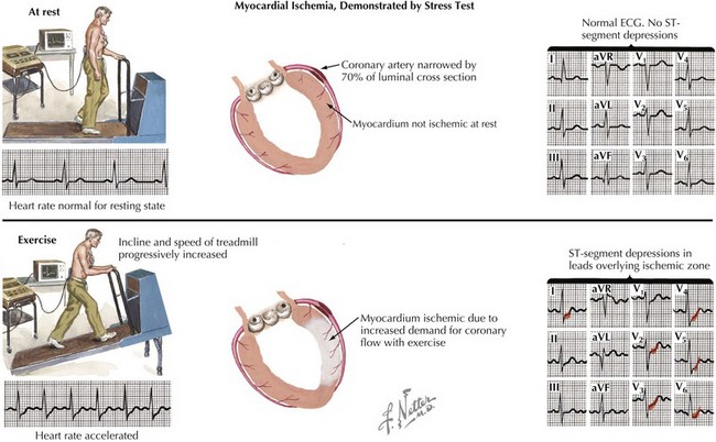
Stress electrocardiography and stress imaging studies are widely used noninvasive procedures that provide important information on cardiac function and the presence of hemodynamically significant coronary artery disease (CAD). The correct use of stress testing is critically important in the cost-effective management of patients with known or suspected CAD. When the most appropriate procedure is performed, it provides important diagnostic and prognostic information that determines the optimal management strategy to be undertaken for that individual. Stress testing is also used in patients with known CAD so as to determine exercise “prescriptions” before cardiac rehabilitation (Fig. 7-1).


Exercise Stress Testing


Exercise stress testing involves subjecting a patient to increasing levels of exercise with continuous electrocardiographic monitoring for myocardial ischemia and arrhythmias. Although the sensitivity and specificity of stress electrocardiography in the detection of CAD are low (in the range of 55% to 75%) compared with more advanced testing (including the use of imaging), stress electrocardiography is widely available, relatively inexpensive, and can provide important prognostic information about the patient. Generally, diagnostic treadmill stress testing is done on patients with a low pre-test likelihood of having CAD. However, exercise stress testing can also be used in patients with known CAD to evaluate the effectiveness of current therapies, to ascertain overall functional capacity, and to determine general prognosis. In children with congenital heart disease, treadmill stress testing can be used to quantify functional capacity.


The sensitivity of exercise stress testing for detecting CAD is proportional to the heart rate (HR) achieved during exercise. Thus, in preparation for the study, patients are usually asked to transiently discontinue medications that affect HR response (e.g., β-blockers or calcium channel blockers). Patients should fast for at least 4 hours before the test. Exercise is done on a treadmill or, alternatively, using a bicycle ergometer. In special circumstances, arm ergometry and isometric hand exercises can be used. There are several different protocols for treadmill stress testing. All of them start exercise at a given rate and incline angle and then gradually increase one or both parameters until an adequate HR and exercise endurance are achieved. Generally, exercise is continued until the patient reaches a target HR of at least 85% of the maximum predicted HR (MPHR) for the patient’s age (220 bpm − age in years ± 10 to 12 bpm). Studies that have correlated ECG changes with CAD generally involve reaching this target HR. Once a patient reaches the target HR, he or she should continue to exercise until fatigued or until signs or symptoms develop. If a patient exceeds a double product (HR × systolic blood pressure) of 25,000 as a secondary target, the test may be considered adequate. If the patient does not attain an exercise level at least equivalent to 5 metabolic equivalents, the study may be considered inadequate. Hemodynamic instability, gross ECG changes, or severe patient symptoms are also indications to terminate the procedure.


At the end of exercise testing, the patient slowly reduces the intensity of exercise. Vigorous exercise results in increased blood flow and pooling in the extremities, and a “step-down” phase (low-level exercise) allows the patient to re-equilibrate before ceasing exercise. After exercise termination, patients are monitored in a supine position until they are no longer tachycardiac (i.e., HR < 100 bpm) if not back to baseline HR. Importantly, if there were any ECG changes or symptoms experienced by the patient during the study, post-test monitoring should be continued with any necessary treatments until these have resolved, even if hemodynamics (HR and blood pressure) have returned to acceptable levels. The post-test monitoring serves to reveal any arrhythmias or ST-segment changes that may develop and be late signs of ischemic disease (Fig. 7-2).


The ECG must be interpreted with certain caveats. Although the standard 12-lead configuration can be used, in many instances a modified 12-lead configuration is substituted. This involves placing limb leads more proximally than is done for a standard ECG (electrodes are placed on the shoulders rather than arms for instance). This change results in ST-segment changes being accentuated and more easily detected during stress. This may also result in a baseline “stress” ECG that differs from a supine ECG done with standard lead placement.


The presence of myocardial ischemia during the test is suggested if previously normal ST segments show flattened or down-sloping depression more than 1 mm below the baseline in three consecutive beats. An important issue concerns ST-segment changes that can occur in some individuals simply because of the increased respiratory rate that accompanies exercise. A pre- or post-stress ECG performed with hyperventilation should be done to allow comparison of ECG changes associated with increased respiratory rate.


The prognostic information obtained from a treadmill stress test is often very useful for deciding on the next diagnostic or therapeutic step for a given patient. Of the several methods used for prognosis following treadmill stress testing, the most widely used is the Duke Treadmill Score. The time of exercise, the presence (or absence) of ST-segment changes during the study, and patient symptoms are used to determine a “score” that correlates with event-free survival.


Bicycle-based studies use a comparable approach to provide similar information. The patient maintains a steady—or, rarely, increasing—pedaling rate over a period of time with regular increases in the intensity required for pedaling. At comparable HRs, a higher level of physiologic stress (reflected by metabolic equivalents) is present in individuals walking on a treadmill than individuals pedaling a bicycle. The data available for comparing these two forms of exercise are quite limited, however. Caution should be used in translating clinical information between forms of exercise.


Contraindications to exercise include unstable coronary syndrome, decompensated heart failure, severe obstructive valvular and hypertrophic cardiomyopathic disease, untreated life-threatening arrhythmias, and advanced atrioventricular block. (Under certain circumstances, exercise testing under rigorously controlled conditions is performed on patients with aortic stenosis to determine their suitability for aortic valve replacement surgery.) Severe baseline hypertension (>220/120 mm Hg) or presence of large arterial aneurysms are also contraindications, as are systemic illnesses such as acute pulmonary embolus and aortic dissection. Exercise studies should be used cautiously in individuals with an implantable cardiac defibrillator, particularly if their underlying ECG shows a prolonged QRS interval (due to an underlying bundle branch block or paced rhythm), because in this circumstance, the defibrillator may “recognize” the rapid HR induced by exercise as ventricular tachycardia. Individuals with an abnormal baseline ECG, particularly with ST-segment abnormalities, should be referred for a stress imaging study, because ECG changes in the setting of an abnormal baseline ECG are far less specific for CAD. Patients with significant left ventricular hypertrophy on their baseline ECG or those taking digoxin have similar limitations for interpretation of ischemia with exercise. Arrhythmias such as uncontrolled atrial fibrillation may also make interpretation of exercise stress ECGs difficult or impossible, and patients with these arrhythmias should be considered for a stress-imaging study.


Cardiac Stress Imaging


Stress-imaging studies combine either treadmill stress testing or an infusion of either dobutamine or a coronary vasodilator (most commonly dipyridamole or adenosine) with imaging of the heart. Imaging can be accomplished by a variety of modalities; those most commonly used are echocardiography or nuclear imaging. MRI has also been used and CT is being studied as a modality for stress imaging. Stress imaging is preferred over treadmill stress testing in several settings: (1) when the ECG is uninterpretable for myocardial ischemia (e.g., left bundle branch block, digoxin effect); (2) when a patient is unable to exercise (but can undergo a pharmacologic stress-imaging study); or (3) when a treadmill stress test is positive for ischemia in a low-risk patient, and correlation by imaging is preferred to cardiac catheterization. Many physicians also prefer stress imaging as a primary approach, rather than ECG-only stress testing, for all patients because of the higher sensitivity and specificity of stress imaging. Even with rapid advances in other modalities, stress imaging remains a highly effective and available modality to evaluate ischemia and function at present, and it is likely that this will be the case in coming years.


Radiotracers


Thallium-201 (201Tl) thallous chloride, a radioactive analogue of potassium, was the most commonly used tracer for myocardial perfusion for several decades. Although its use has declined with the advent of technetium-99m (99mTc)–based agents, 201Tl continues to be useful as part of dual-isotope protocols and in viability imaging. Its relatively low energy results in images that lack resolution. However, the higher myocardial extraction fraction of 201Tl compared with 99mTc-based agents has resulted in its continued use.


The two most commonly used 99mTc-based MPI agents are 99mTc-sestamibi (MIBI) and 99mTc-tetrofosmin. Images obtained with the two agents are comparable and have higher resolution than images obtained using 201Tl for cardiac imaging. MIBI demonstrates a slightly higher extraction fraction than tetrofosmin and is therefore more commonly used, although the use of MIBI results in a slightly higher radiation dose to the patient compared with tetrofosmin. A previously used 99mTc-based agent, teboroxime, demonstrated a substantially higher extraction fraction than the aforementioned agents, but its rapid washout from the myocardium limited its clinical utility. Teboroxime is no longer marketed in the United States.


PET radiopharmaceuticals utilize positron-emitting radionuclides to create images. Rubidium-82 (82Rb) chloride is a positron-emitting potassium analogue. It has the lowest extraction fraction of the available PET radiopharmaceuticals (~60%). This extraction fraction is still higher than that of either sestamibi or tetrofosmin. The half-life of 82Rb is very short—approximately 75 seconds. There are benefits and limitations for the use of 82Rb given its very short half-life. The short half-life essentially precludes use of 82Rb for exercise stress imaging. However, it facilitates obtaining images when the patient is truly at the peak of performance induced by pharmacologic stress. For this reason, 82Rb images can be used to accurately assess cardiac reserve—as defined as the difference between left ventricular ejection fraction (EF) at rest and at peak stress. The short half-life of 82Rb also facilitates obtaining pharmacologic stress and resting images in a relatively short period of time.


82Rb has a lower intrinsic spatial resolution than the other PET agents but is still far better than the SPECT tracers. Although a cyclotron is not necessary to generate 82Rb, the generator system used is quite expensive and, for this reason, 82Rb PET imaging is only available at some centers.


Other tracers are used for PET imaging, but none are used for cardiac imaging as commonly as 82Rb. Nitrogen-13 ammonia ([13N]NH3) has a high extraction fraction (approximately 83%) and a 10-minute half-life. It can be used for exercise-nuclear imaging. Oxygen-15 ([15O]H2O) water is short-lived (half-life of 2 minutes) and possesses a very high extraction fraction of approximately 95%. However, its freely diffusible nature means that 15O is distributed into tissues adjacent to the myocardium, including the lungs and cardiac blood pool. For this reason, imaging is complicated, requiring sophisticated background subtraction techniques. Although both 13N and 15O have higher intrinsic spatial resolution than 82Rb, they require generation in a cyclotron. Their short half-lives mean that these isotopes can only be used in facilities with an on-site cyclotron. For most institutions performing PET-myocardial imaging studies, 82Rb is preferred for this logistic reason.


Newer fluorine-18 (18F)–labeled perfusion tracers that would allow exercise imaging and do not require an on-site cyclotron are being developed and studied. The 18F tracers have a very high extraction fraction, making them physiologically attractive in the assessment of CAD.


Jun 12, 2016 | Posted by in CARDIOLOGY | Comments Off on Stress Testing and Nuclear Imaging

Full access? Get Clinical Tree

Get Clinical Tree app for offline access