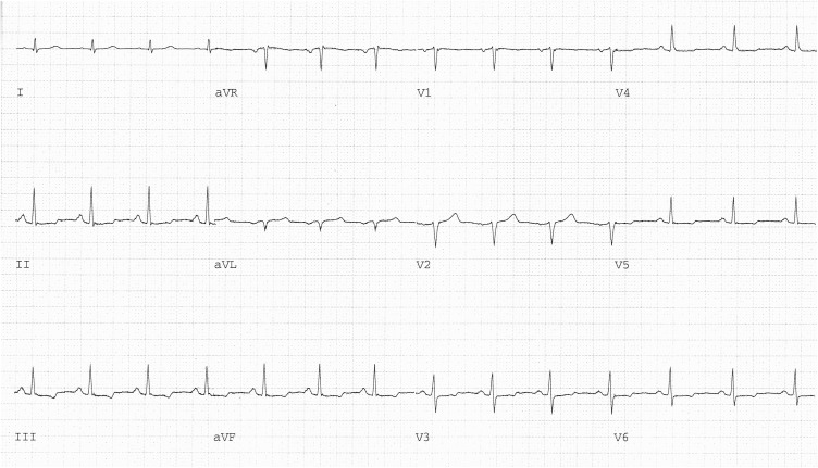
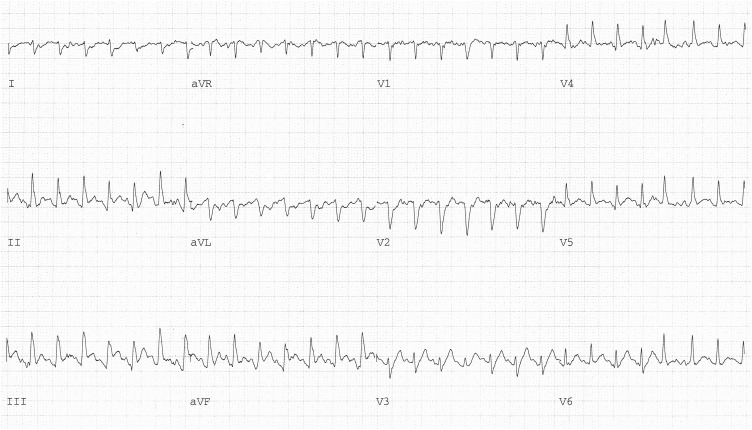
A 53-year-old man, a former smoker with a history of hyperlipidemia, came to the hospital because of 4 weeks of chest pain occurring with walking. He underwent a treadmill exercise test using the standard Bruce protocol, 12-lead electrocardiography, and two-dimensional echocardiography. The resting electrocardiogram showed sinus rhythm at a rate of 79 beats/min and nonspecific ST-T changes in the inferolateral leads ( Figure 1 ). The resting echocardiogram showed normal left ventricular function. The patient exercised for 10 minutes and 30 seconds and stopped because of the onset of chest pain and the development of ST-segment elevation in leads II, III, and aVF with reciprocal ST-segment depression in leads I and aVL. In addition, the QRS axis had shifted rightward, and Q waves had appeared in leads II, III, and aVF ( Figure 2 ). The echocardiogram performed immediately after exercise showed severe inferoposterior hypokinesis. All these changes soon disappeared spontaneously.
ST-segment elevation occurring with exercise is common in patients with previous Q-wave myocardial infarcts and usually occurs in the leads with the Q waves and the corresponding left ventricular dyskinesis or akinesis. Although such exercise-induced ST-segment elevation may be due to ischemia in residual viable myocardium, in most cases, passive wall motion abnormality is probably the cause.
In patients, such as ours, without infarction, ST-segment elevation during exercise is rare, implies severe transmural ischemia, and can be due to high-grade fixed coronary arterial narrowing or coronary arterial spasm. The ST elevation occurs in leads corresponding to the coronary artery with the severe fixed narrowing or the spasm. This is unlike the more common exercise-induced ST-segment depression, which often indicates myocardial ischemia but cannot localize it accurately. Exercise-induced Q waves also are uncommon, may be due to myocardial ischemia, but are neither specific nor sensitive markers for coronary disease.
Our patient’s inferior ST-segment elevation and inferoposterior wall-motion abnormalities that appeared with exercise predicted his coronary arteriographic findings: a >95% narrowing in the midportion of the right coronary artery, which was successfully stented. Because of a 60% stenosis in the left anterior descending coronary artery, the patient returned for a repeat treadmill exercise test to access the lesion’s significance. The second exercise test was entirely normal. The patient remained alive and well at the last follow-up 10 years later.
Disclosures
The authors have no conflicts of interest to disclose.
See page 1949 for disclosure information.
Stay updated, free articles. Join our Telegram channel

Full access? Get Clinical Tree


