Squamous Cell, Basaloid, and Lymphoepithelial-Like Carcinoma


Squamous Cell, Basaloid, and Lymphoepithelial-Like Carcinoma


Andre L. Moreira, M.D.

Fabio R. Tavora, M.D., Ph.D.

Allen P. Burke, M.D.


Invasive Squamous Cell Carcinoma


Epidemiology

Between 1977 and 1986, squamous cell carcinoma accounted for more than 30% of lung cancers, a rate that has decreased to <20% because of a decrease in cigarette smoking.1 Approximately 224,000 new lung cancers (of all types) were predicted to occur in the United States in 2014,2 indicating about 45,000 that are of the squamous type. Although there is a nearly equal gender distribution for all lung cancers,2 there is still a male predominance in most series of squamous carcinomas of the lung.3


TABLE 78.1 Rate of Positivity, Pulmonary Squamous Cell Carcinomas, Selected Immunohistochemical Markers


Antibody


Resected Tumors


TMA Samples


Peripheral Tumors (TMA)


Central Tumors (TMA)


p63


100%2,3,4,5


100%6


94%7


95%12


71%1


65%1


p40 (δNp63)a


100%d


100%6


81%7


Cytokeratin 7


20%-60%2,3


40%1


4.7%1


TTF-1


0%2,3


3% (focal)4


06


0%12


2%12


5%1


0%1


Napsin-A


0%2


2%12


Cytokeratin 5/6


73%-100%2,3,4


53%12


85%1


90%1


34βE12 (CK903)


100%2,4


p16


All tumors


10%9; 21%13; 63%8


HPV+b


71%8-100%9,11


HPV-


010; 6%9; 18%11; 21%13; 62%8


p53


All tumors


53%8


HPV+


76%8


HPV-


50%8


1. Saijo et al. (2006).10

2. Mukhopadhyay and Katzenstein (2011).11

3. Pelosi et al. (2011).12

4. Rekhtman et al. (2011).13

5. Bishop et al. (2012).14

6. Nonaka (2012).15

7. Ao et al. (2014).16

8. Fan et al. (2015) Cytoplasmic OR nuclear staining.17

9. Gatta et al. (2012) Nuclear AND cytoplasmic staining.18

10. van Boerdink Nuclear OR cytoplasmic >70% of tumor.7

11. Yanagawa et al. (2013) Nuclear only, > 10%.6

12. Whithaus et al. (2012); TTF-1 stained 2% of squamous carcinoma using one antibody (Leica) and 0% using another (Dao).19

13. Bishop et al. (2012); strong and diffuse cytoplasmic and nuclear staining in >70% of tumor cells.14


TMA, tissue microarray.


a Preferred because of greatest specificity for squamous differentiation.

b By in situ hybridization and/or polymerase chain reaction, primarily metastatic tumors.


Risk Factors

Similar to small cell carcinoma, squamous cell carcinoma is highly related to degree of exposure to cigarettes, especially to tar in the smoke.4 A man who smokes more than 30 cigarettes a day is 103.5 times more likely to develop squamous carcinoma of the lung than is a nonsmoker.5 Occupational agents that increase the likelihood of lung cancer, including squamous carcinoma, include asbestos and arsenic.4 The association between human papillomavirus infection and squamous lung carcinoma is weak, at least in Western populations.6,7


Radiologic Findings

Although two-thirds of all squamous carcinomas are central, squamous cell carcinomas that are surgically resected are slightly more likely to be peripheral.8,9 On computed tomography scans, they are less often spiculated, less often have internal air bronchograms, and are more likely to have heterogeneous enhancement and cavitation, as compared to pulmonary adenocarcinomas.9


Gross Findings and Staging

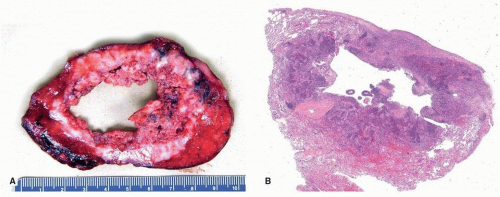
Central squamous carcinomas usually arise in a main or lobar bronchus (Table 78.1). These tumors may form intraluminal polypoid masses or endobronchial papillary tumors (Fig. 78.1).20 Uncommonly, they spread superficially and are limited to the bronchial wall, in which case they are classified as T1a, regardless of size and whether or not they extend to the main bronchus.21 Squamous cell carcinoma typically infiltrates adjacent structures through direct invasion. On cut surface, they are firm and gritty, and there may be focal carbon pigment deposits in the center. Cavitation occurs in about 20% of tumors, typically those that attain a large size (Fig. 78.2). Postobstructive changes, including endogenous lipid pneumonia, are common in central lesions.


Microscopic Findings

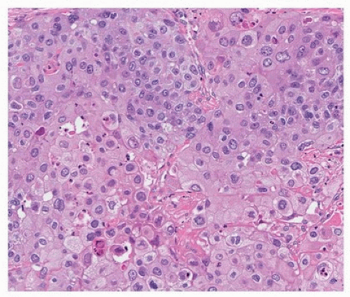
Well-differentiated squamous carcinomas typically have intracellular bridges and foci of keratinization and form sheets of cells
without glandular structures or mucin vacuoles (Figs. 78.3 and 78.4). Nonkeratinizing, poorly differentiated tumors may have a basaloid appearance (see below) or resemble adenocarcinomas with a solid growth pattern, in which case immunohistochemical stains are necessary for a definitive diagnosis. Proximal tumors tend to have a papillary or endophytic growth pattern (Fig. 78.5). Peripheral tumors grow in two overlapping patterns: one that respects alveolar architecture, filling alveolar spaces (Fig. 78.6), and one that is accompanied by fibrosis, with a pushing, destructive edge (Fig. 78.7).8,22 Peripheral tumors are more likely to have vascular (Fig. 78.8) and pleural invasion and less likely to be associated with squamous metaplasia in adjacent bronchi than proximal tumors.10 There may be large areas of cytoplasmic clearing due to intracytoplasmic glycogen (Fig. 78.9), a nonspecific finding that is common in both squamous and adenocarcinoma. Squamous carcinomas may grow along alveolar walls, a pattern that is often confused with adenosquamous carcinoma. This growth pattern has been termed “endoalveolar growth” or “invasion along alveolar walls” and is seen in up to 20% of squamous carcinomas.23,24
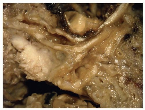


FIGURE 78.1 ▲ Squamous cell carcinoma. The tumor distorts the bronchial surface and invades the adjacent lung.


FIGURE 78.2 ▲ Squamous cell carcinoma. Approximately 20% of tumors, especially those that are central, form cavitary masses. A. Gross photograph. B. Corresponding histologic section.


FIGURE 78.3 ▲ Squamous cell carcinoma. Tumor forms cohesive sheets with scattered dyskeratotic cells.

Pathologic studies of early squamous carcinomas that are occult on chest x-ray and detected by screening show that they progress from in situ lesions to mucosal, muscular, cartilaginous, and extrabronchial invasion.25 Infiltration of the bronchial wall is of two types: one extensive bronchiolar spread without extending through the cartilage and the other penetrating deeply into the wall, presumably advancing within a short period.25

There is no standardized grading of squamous carcinomas, although the proportion of tumor with keratinization or intracellular bridges is a useful grading tool (Fig. 78.10). Well-differentiated tumors show more than 50% keratinization with intracellular bridges; moderately differentiated tumors have less than one-half; and poorly differentiated tumors have minimal if any keratinization.11


Immunohistochemical Stains

As in other sites, a variety of markers (p63, p40, cytokeratin 5/6, cytokeratin 903/34βE12) are used to identify squamous differentiation in lung carcinomas (Table 78.1). Cytokeratin 903, although highly sensitive,

lacks specificity (Table 78.2). δNp63 (p40) is of similar sensitivity as p63, but more specific, as it is less likely expressed in adenocarcinomas and neuroendocrine carcinomas than p63.26 For this reason, it has largely replaced p40 in distinguishing poorly differentiated squamous from adenocarcinomas of the lung. Cytokeratin 5/6 is nearly as sensitive and specific as p40 and is often used in panels that include other markers in the distinction between squamous and other carcinomas of the lung.11,12,13

Only gold members can continue reading. Log In or Register to continue

Stay updated, free articles. Join our Telegram channel

Aug 19, 2016 | Posted by in CARDIOLOGY | Comments Off on Squamous Cell, Basaloid, and Lymphoepithelial-Like Carcinoma

Full access? Get Clinical Tree

Get Clinical Tree app for offline access