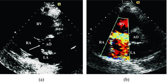
Echocardiogram revealed that left ventricle (LV) and RV were normal in size and systolic function. A PSA view with color Doppler shows a ruptured aneurysm of the noncoronary sinus of Valsalva and the high-velocity flow through the ruptured noncoronary sinus aneurysm into right atrium (RA; Figure 1.2 and Videoclip 1.2).
Figure 1.2 (a) Parasternal short-axis view shows ruptured aneurysm (arrow) of the noncoronary sinus of Valsalva. (b) Parasternal short-axis view with color Doppler illustrates the high-velocity flow through the ruptured noncoronary sinus aneurysm into right atrium in case 2. AO, aorta; LA, left atrium; RA, right atrium; RV, right ventricle.
Discussion
Stay updated, free articles. Join our Telegram channel

Full access? Get Clinical Tree


