Chapter contents
6.1 Introduction 72
6.2 The simple pulse parameters 73
6.3 Rate 73
6.4 CM pulses defined by rate 78
6.5 Rhythm 83
6.6 CM pulses defined by rhythm 88
6.7 Depth 91
6.8 CM pulse qualities defined by level of depth 94
6.9 Length (longitude) 100
6.10 CM pulse defined by length 101
6.11 Width (latitude) 103
6.12 CM pulse qualities defined by arterial width 110
6.13 Summary 112
Pulse diagnosis is often complicated in the clinical context because pulse manifestations as they present in the radial artery do not present as nice discrete ‘images’ as posited in the literature. Indeed, expectations of always feeling a classical pulse quality are misplaced and will lead to difficulties and frustration on the part of the pulse taker. Rather, it should be expected that the pulse may ordinarily present as no recognisable traditional pulse quality. This is to be expected, as each person will not always respond the same way to the same illness: a person’s constitution and the relative strength of their Qi and blood means the body responds differently. Other factors such as lifestyle (work, exercise, dietary) and life history only add further variability.
There may be instances when the pulse taker assesses the pulse when the immune system is just responding to a pathogenic agent so the pulse is felt while it is moving from the individual’s normal or healthy pulse to the pulse that reflects the nature of the pathogenic illness and the body’s response to that illness. In this situation, the pulse is not yet fully formed and may also not fit nicely into the distinctive CM pulse qualities described in the literature. Alternatively, the body’s response to the illness is such that the pulse quickly forms a recognisable quality and it is over time as the body’s energies are depleted or the pathogen mutates that the pulse quality may assume a less recognisable form.
In this sense, the pulse can be thought of in terms of a continuum (Fig. 6.1). The idea of the pulse as a continuum is purposely used to denote that the pulse is not a static sign, nor is the body a static organism. In this sense, there are no ‘absolutes’ as to how the pulse should be, it always just is . It is reflective of the relative degree of the body’s ability to maintain normal healthy function or homeostasis in response to the constant demands of living. In terms of illness or dysfunction, the idea of the continuum refers to the changing flux of the pulse between two points of reference. At one reference point are those pulses reflecting health, usually assumed to be the individual’s usual pulse state; at the other point of reference are those pulses that reflect illness or dysfunction — the potential end manifestation of the pulse flux if no treatment intervention is provided to counter the pathogen/dysfunction.
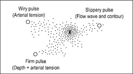 |
| Figure 6.1Schematic of the pulse in a continuum. The pulse formation illustrated has aspects of the Firm pulse, Slippery pulse and Floating pulse and so does not discretely fit into any of these pulse categories as they are defined within the literature. |
Sometimes the pulse palpated is in flux on a continuum between two pulse types that both reflect illness or dysfunction; there may be a worsening or improvement in an illness, but not necessarily resolution of the illness. Chronic inflammatory conditions such as arthritis are examples of diseases in which there are acute periods of inflammation interspaced with periods of generalised symptoms. In this respect, certain pulse qualities develop slowly, progressing through stages in response to physiologic changes associated with disease progression or resolution. Such a process is comparable to that described by Katz (2000) in relation to the problems of defining heart failure.
… this condition is not a disease but instead represents the final common pathway by which a number of disorders damage the heart so as to cause disability and premature death. These disorders include coronary disease, hypertension, valvular disorders, and a diverse group of heart muscle diseases referred to as cardiomyopathies. Furthermore, because this syndrome establishes a number of vicious cycles, heart failure begets more heart failure. (p. 7)
The description is particularly pertinent to pulse diagnosis. In this respect, pulses reflecting chronic illness and dysfunction arising from the consumption of vital substances occur over time. Some of the traditional pulse qualities are reflective of this chronological progression of illness and will only occur as an end result of a long disease process. Other pulses occur only in an acute situation. Some other pulse qualities occur only when fundamental substances of Qi, Blood and Fluids are abundant; when these are depleted, then those pulse qualities will not occur.
Potentially there is an immense range of combinations of the pulse parameters; it is the interaction of these and their various manifestations that produces unique pulses that do not fit easily into the discrete pulse images presented in the literature. In such instances, recording and noting the pulse in terms of its constituent parameters allows important diagnostic information to be available for use, rather than lost within an incorrect application of a pulse quality name.
6.1. Introduction
The term pulse parameters refers to the fundamental variable characteristics that contribute to the formation of the radial arterial pulse. The 27 traditional CM pulse qualities form when there are specific changes in these parameters. A traditional pulse quality can form when there is a change in a single parameter only; other traditional pulse qualities form when there are changes in several parameters. In the pulse parameter system of radial pulse diagnosis, we have identified 9 pulse parameters that determine the 27 CM pulse qualities:
• Rate
• Rhythm
• Arterial width
• Depth
• Length
• Arterial tension
• Ease of occlusion
• Force
• Pulse contour.
6.1.1. Parameters and the arrangement of the CM pulse qualities
In Chapter 6 and 7 we present each of the 27 specific CM pulse qualities in conjunction with their relevant defining parameter(s). A pulse’s defining parameter refers to the variable change in the presentation of the pulse that is most apparent. For example, the Rapid pulse is defined by an increase in the frequency of pulsations. Consequently, the Rapid pulse’s defining parameter is pulse rate.
Where distinct changes in two or more parameters are involved in the formation of the CM pulse quality, we have categorised the CM pulse quality by using the parameter that plays the most important role in its formation. For example, the Skipping pulse features interruptions to the pulse’s regular rhythm and is accompanied by an increase in pulse rate. Yet it is the rhythm changes that are its most defining parameter and it is consequently categorised under the rhythm parameter.
However, this categorising of the pulse qualities is by no means the only way to classify them. Some of the more complex CM pulse qualities presented in Chapter 7 could be classified under more than one parameter. Categorising a pulse based on parameters is flexible.
Depending on the number of parameters involved in a pulse’s formation, the traditional CM pulse qualities can be additionally categorised as simple or complex. Simple refers to pulse qualities that are defined by a change in one parameter only, like the Rapid pulse. Complex refers to pulse qualities that are defined by a change in two or more parameters, such as the Firm pulse. The terms ‘simple’ and ‘complex’ can equally apply to the pulse parameters themselves. For example, pulse rate is a simple pulse parameter as it is characterised only by the pulse frequency. In contrast, pulse force is a complex pulse parameter because it depends on a range of variables such as the strength of cardiac contraction, blood volume and the tensile compliance of the arterial wall. It is how these variables combine that determine the force of the pulse.
In addition to presenting the traditional 27 pulse qualities with their relevant defining parameters, we also present the pulses according to the complexity of their defining parameter. Each of the parameters are defined, assessment techniques are detailed and the related CM pulse qualities noted along with their clinical significance. Simple pulse parameters and the related CM pulse qualities are presented in this chapter. Complex pulse parameters and related CM pulse qualities are presented in Chapter 7.
6.2. The simple pulse parameters
The simple pulse parameters covered in this chapter are:
• Rate
• Rhythm
• Depth
• Length
• Width.
Related changes in these five parameters are associated with 12 of the 27 traditional CM pulse qualities. The CM pulse qualities associated with the simple pulse parameters are:
• The Slow, Rapid and Moderate pulses (defined by rate)
• The Skipping, Bound and Intermittent pulses (defined by rhythm and accompanied by changes in rate)
• The Sinking, Floating and Hidden pulses (defined by level of depth)
• The Long and Short pulses (defined by length)
• The Fine pulse (defined by its width).
These pulses can be classified as simple pulse qualities because they are generally defined by a change in a single parameter. Exceptions are the Bound pulse and Skipping pulse, which form as a result of changes in two parameters, rhythm and rate. However, the Bound pulse and Skipping pulse are still considered ‘simple’ pulse qualities as they are defined by two of the most objectively evaluated parameters.
These five parameters are also defined as simple because they are relatively easy to assess in a clinical context. Indeed, these five parameters are deemed the least subjective of the nine parameters, involving the evaluation of distinctive physiological characteristics of the artery and the pulse wave. For example, manual palpation of the radial arterial diameter is assessed for the parameter of width. Pulse length is assessed by the presence or absence of pulsations at Cun, Guan and Chi pulse positions and beyond these positions. The parameter of depth entails an appraisal of the levels of depth where pulsations are relatively strongest. The rate and rhythm of the pulse can be calculated with standardised formulae of beats per minute in the case of rate, and comparison of the length of intervals between beats for rhythm. Pulse rate and rhythm can also be accurately assessed using electronic devices such as electrocardiography, and arterial width can be measured using Doppler ultrasound.
In a biomedical context, the three parameters of rate, rhythm and width are used primarily to provide information about the functional performance of the cardiovascular system, particularly the heart. The parameters of depth and length are not extensively used in the biomedical diagnostic sense, but are used as an indicator of the circulatory system’s integrity. This is best seen in acute traumatic injury of the limbs in which circulation may be compromised through swelling and fractures causing arterial occlusion, and also occurs in chronic conditions in which arteries narrow and blood flow is impeded. Palpating the length of the artery is useful in identifying the point of arterial occlusion, and pulse depth is used to assess the strength of blood flow and pressure in the vessel.
6.3. Rate
Three traditional CM pulse qualities are associated with the pulse rate parameter:
There are clear guidelines in the literature that detail when to interpret pulse rate measures outside the normal stated pulse rate range as healthy. This often depends on a range of variables including gender, age and exercise. Therefore, in clinical practise it is important to record the pulse rate every time you palpate an individual’s pulse, to establish a normal baseline measure for their particular pulse rate.
6.3.1. Pulse rate and its measurement
The pulse wave that we ultimately feel in the radial artery originates in the heart, due to the rhythmic contraction (systole) and relaxation (diastole) of the heart’s left ventricle as it pumps blood throughout the body. Each systole and the following diastole is known as one cardiac cycle . Therefore, pulse rate is an expression of the heart rate, describing the number of times the cardiac cycle occurs each minute.
Assessment of pulse rate involves noting the presence of pulsations and the frequency with which these occur in one minute. Although pulse rate is expressed in beats per minute, it isn’t always necessary to assess the pulse frequency for a full minute. For example, one method for assessing pulse rate involves counting the number of beats for 15 seconds then multiplying this value by 4, or counting the number of beats for 30 seconds, then multiplying this value by 2, to obtain a measure of pulse rate in beats per minute (bpm).
The assessment method used for obtaining a measure of pulse rate should be repeated at least twice and the findings averaged. This is useful in two ways. Firstly, it assists in determining the accuracy of the value obtained for the pulse rate. Secondly, by obtaining a second measure of pulse rate, any transient changes affecting heart rate are more readily identified by noting any large variations between the two measured values. This also increases the reliability of pulse rate assessment. (See Box 6.1 for further considerations when interpreting pulse rate diagnostically.)
Box 6.1
Questions to consider when assessing pulse rate
• Is this a first-time patient?
• Has the patient been hurrying to the appointment?
• Are they nervous or stressed?
• Does the patient exercise regularly? What type of exercise do they do?
• Have they exercised recently before their appointment?
• Have they taken any medications/supplements?
• Has the patient consumed tea or coffee recently? Are there any other dietary sources of caffeine, for example carbonated or energy drinks, herbal supplements?
• Is the pulse wave rising slow, normal or fast?
• Is there aversion to heat or cold?
• Is a fever present?
• What is the weather like?
6.3.1.1. Pulse rate versus heart rate
The terms pulse rate and heart rate are often used interchangeably to mean the same thing. This is based on the assumption that since heart contraction produces the pulse wave, all pulse movements felt at the radial artery should correspond to the same number of heart contractions. However, there are certain biomedical conditions affecting the heart and arterial structures in which two distinct pulse waves can be felt for every heart contraction. Sometimes the heart contraction can be weak and the pulse wave cannot be felt in the radial artery. In other cases the pulse is being occluded, as in thoracic outlet syndrome. It should be noted then that in certain circumstances, the pulse rate and the heart rate do not correspond.
6.3.2. Normal pulse rate
The average resting pulse rate in a healthy adult is about 70–80 bpm, but this ‘normal’ range is usually extended to 60–90 bpm. Normal pulse rate will also vary depending on the level of physical activity. In a healthy adult, it can decrease to 40 bpm during sleep. At the other extreme, pulse rates of up to 180 bpm may occur during intense exercise (Epstein et al 1992: p. 7.9).
6.3.3. Variables affecting pulse rate
Of all the pulse parameters, pulse rate is the most variable. It is easily affected by several factors, including:
• Gender
• Age
• Exercise
• Medications
• Body temperature
• Emotions.
6.3.3.1. Gender and pulse rate
Estes (2006: p. 253) asserts that women have a slightly faster resting pulse rate than men:
• Women: 72–80 bpm (average 75)
• Men: 64–72 bpm (average 68)
Gender-related pulse rate differences are thought to result from the relative size of the heart. Men have relatively larger hearts than women resulting in a greater proportion of blood being pumped through the arterial system with each cardiac contraction. In women, if the heart is relatively smaller, to maintain the same blood volume movement as men the heart rate needs to increase. (This gender difference is a generalisation, as commonly both men and women will have normal resting heart rates greater than or less than the ranges listed.)
6.3.3.2. Age and heart rate
There is an inverse relationship between heart rate and age; heart rate decreases slightly with increasing age. This is a result of decreasing responsiveness of the β-adrenergic receptors in the cardiac cells to chemical stimulus, combined with decreasing sinoatrial node automaticity (McCance & Huether 2006) (see the description of the intrinsic conduction system in section 6.4.5).
From a CM perspective this correlates to Yang Qi decreasing with age. Children and infants, considered to have more Yang Qi intrinsically, have a significantly higher heart rate, with newborns likely to have a heart rate of 140 bpm. Resting heart rate for a fetus can be as high as 140–160 bpm (Marieb 2001: p. 708). Table 6.1 lists a range of normal heart rate ranges for different age groups.
| Age (years) | Range (bpm) | Average (bpm) |
|---|---|---|
| Newborn | 100–170 | 140 |
| 1 | 80–160 | 120 |
| 3 | 80–120 | 110 |
| 6 | 70–115 | 100 |
| 10 | 70–110 | 90 |
| 14 | 60–110 | 85–90 |
| Adult | 60–100 | 72 |
| Adult men | 64–72 | 68 |
| Adult women | 72–80 | 75 |
The relationship between age and heart rate is clinically reflected in the diagnostic interpretation of the resting heart rate. For example, a slow heart rate occurring in a younger person, when not associated with any form of aerobic training, is seen as a poor sign of health. In older people, Perk et al (2003) found a strong correlation between heart rate and all-cause mortality in elderly women in a study examining the relationship between heart rate and mortality in the elderly (average age of subjects was 70 years). Although there was a similar trend for elderly men, this was not statistically significant. Perk et al (2003) found that women with a heart rate greater than 77 bpm had three times the mortality rate of those whose heart rates were less than 77 bpm (r = 0.25, P = 0.0003). This means that a relatively fast resting heart rate (that is, on the higher side of the normal range) in an older person is a mortality risk sign and can be viewed as a sign of poor health (Perk et al 2003). In a CM context, this probably reflects vacuity of the heart associated with depletion of essential substances, notably Qi.
6.3.3.3. Exercise and heart rate
Like any other muscle in the body, the more exercise the heart receives the better toned it becomes and therefore the more efficient in moving blood. This is particularly noticeable in individuals who undertake endurance training and consequently have a slower than normal resting heart rate (50–60 bpm) with elite athletes resting heart capable of falling below 50 bpm (McCance & Huether 2006: p. 1048). A slower heart rate in this situation results from:
• Increased vagal stimulation (which slows the heart) and decreased sympathetic stimulation
• Increase venous return of blood to the heart due to lowered peripheral resistance leading to increased stroke volume.
Stroke volume (SV) is the amount of blood pumped out by a ventricle with each heartbeat. As the heart rate (HR) slows down, this allows longer ventricular filling, which in turn increases stroke volume. The relationship between SV and HR is termed cardiac output (CO), defined as the amount of blood that is pumped out by each ventricle in one minute and dependent on the heart rate and stroke volume:

Therefore, increased SV helps to compensate for a slower rate to maintain CO.
When cardiac fitness is poor, the heart functions less efficiently and so SV is diminished, with less blood being ejected from the heart with each cardiac contraction. In this situation, the HR increases to maintain CO. In individuals who exercise little, or whose cardiac muscle tone is poor, their normal resting heart rate is consequently raised.
6.3.3.4. Medication, herbal supplements and heart rate
There are many medications and herbal supplements that affect heart rate, and appropriate texts should be referenced for further information. Many of the routinely used over-the-counter medications such as cold and flu medications contain compounds such as pseudoephedrine (a decongestant) that may cause heart and pulse rate to increase. Common asthma-relieving medications such as salbutamol, found in bronchodilators, are β-adrenergic agonists which may cause an increased heart rate or tachycardia in some patients. Many herbal medicines and supplements such as guarana (Paullinia cupana), aconite (Fu zi, Aconitum carmichaeli) and ephedra (Ma Huang, Ephedra sinica) have stimulant effects on the heart and pulse rate. Nicotine also has a stimulatory effect on pulse rate (while cannabis has a primary effect on the pulse contour). Digitalis and other drugs such as beta-blockers, which block the β-adrenoreceptors and prevent stimulating chemicals from attaching, may slow down the heart rate.
6.3.3.5. Body temperature and heart rate
An increase in body temperature results in increased heart rate because of a rise in the metabolic rate of cardiac cells. This is commonly seen with fever. Conversely, a decrease in body temperature can slow down heart rate and contractility. Individuals exposed to cold and suffering hypothermia routinely present with slowed heart rates. In a CM context, cold is seen as retarding the body’s Yang Qi and so the pulse slows.
Generally, environmental conditions associated with the seasons similarly affect the pulse. In summer, the pulse is felt strong, relatively superficial and slightly fast, and this is seen as a ‘normal’ response to the environment rather than a pulse representing pathology. Cold weather or seasonal conditions can cause the arterial tension to increase, and pulse width may decrease in an attempt to maintain body warmth. The classical CM literature also described the pulse as being deeper in winter.
6.3.3.6. Emotions such as fear, excitement, anxiety or stress
Extremes of emotion may cause changes in heart rate. Anxiety may cause tachycardia, commonly experienced as a ‘panic attack’. Depression may affect the Shen, thereby affecting the heart’s control of blood and the vessels. Stress stimulates the sympathetic nervous system increasing production of epinephrine (adrenaline) and elevating body heat as a consequence of increased metabolic activity (Estes 2006: p. 255). As noted previously, increase in body temperature can increase heart rate.
6.3.3.7. Pregnancy and heart rate
Early in the first trimester of pregnancy a number of hemodynamic changes take place in order to meet the demands of the growing foetus. Increased cardiac output is due to an increase in both heart rate and stroke volume. The increase in heart rate occurs as early as 4 weeks after conception and increases on average by about 15 bpm (Stables & Rankin 2005: p. 233).
6.3.3.8. Other causes affecting heart rate
There are a number of factors additionally associated with changes in the pulse rate parameter and which are always seen as a pathological response. These include (but are not limited to):
• Decreased potassium (hypokalemia), which delays ventricular repolarisation and may have varying effects on rate; bradycardia — a slow pulse, atrioventricular block or paroxysmal atrial tachycardia (McCance & Huether 2006: p. 105).
• Inhibition of the vagus nerve can cause tachycardia (McCance & Huether 2006: p. 1049).
• Shock and hypovolemia (low blood pressure) due to blood loss, plasma loss or interstitial fluid loss. Initial compensatory mechanisms include increases in heart rate and systemic vascular resistance to elevate cardiac output, by release of catecholamines by the adrenals (McCance & Huether 2006: p. 1628).
• Thyroid hormones cause changes in heart rate. Hyperactivity or hypoactivity of the thyroid affects metabolism and core body temperature (McCance & Huether 2006: pp. 692-5).
6.3.4. Regulation of heart rate: CM perspective
The rate of the pulse provides a general indication of the functional activity of Yang Qi in the body (Box 6.2). Yang Qi is seen as a motive force, giving rise to and ensuring the regularity of the movement of both Qi and blood:
Box 6.2
Traditional CM method of pulse rate assessment
The objective evaluation of heart rate with a timepiece such as a watch was not available at the time the CM classics were written. Instead, a method was devised for the purpose of evaluating whether the pulse rate was faster or slower than it should be. This method was based on the number of beats per complete respiration cycle of the patient (one inhalation and one exhalation) or the respiratory rate of the practitioner, depending on the CM literature reviewed. The following quote from the Su Wen Nei Jing describes this approach:
In man,
during one exhalation, the vessels exhibit two movements.
During one inhalation, the vessels exhibit two movements too.
Exhalation and inhalation constitute one standard breathing period.
If the vessels exhibit five movements,
this is an intercalation [of a fifth movement] because of a deep breathing.
That is called a ‘normal person’.
Su Wen
From the pulse literature, the normal pulse rate per complete respiration cycle breath should be 4–5 pulse beats (see Table 6.2 for age-related respiratory rates). A later expansion of the Su Wen passage by a commentator suggested that a patient’s condition should be assessed by making a comparison with the pulse frequency of someone who is not ill, such as the healthy physician.
Disadvantages of the traditional method of pulse assessment
The obvious disadvantages of the respiratory method include:
• The assumption that the practitioner is in good health and therefore has a ‘normal’ rate of respiration to provide a reliable baseline comparison. This is not always the case.
• Lack of agreement between literature sources relating to the use of the respiratory rate of the practitioner or the patient as the baseline comparison.
• If using the patient’s respiratory rate, it may be difficult to observe the complete respiratory cycle, as it is not always easy to see inspiration and expiration clearly, particularly if the patient’s breathing is shallow or irregular.
That which is quiet is Yin; that which moves is Yang
That which is retarded is Yin; that which is accelerated is Yang.
Su Wen 7
The pulse rate is affected when the Yang Qi is affected. In this context, changes in pulse rate occur when Yang Qi is affected by:
• External factors causing heart rate to increase or decrease depending on whether the cause is of a hot or cold nature. External factors include dietary and pathogenic causes.
• Internal factors causing heart rate to become hyperactive (often due to Yin vacuity so the Yang is no longer controlled) or hypoactive (through Yang Qi vacuity, in which the Yang Qi is no longer sufficient to move the heart and blood) at its customary rate.
Additional changes in the pulse parameters of force (generally increased in replete patterns and decreased in a vacuous patterns) and depth are used to further differentiate the pulse rate changes and the causes. Information obtained from assessment of the pulse rate parameter is always used with information obtained from the assessment of other pulse parameters. Pulse rate assessment alone does not supply sufficient information to identify the location (internal or external) or the nature (vacuity or replete) of disease/dysfunction.
6.3.5. Regulation of heart rate: biomedical perspective
From a biomedical perspective, there are three regulatory mechanisms which control heart rate (the following information is from McCance & Huether 2006):
• Intrinsic conduction system: The fundamental rhythm of the heart is set by the sinoatrial (SA) node, a small area of cells that function as a pacemaker due to their spontaneous electrical nature. They continuously depolarise and initiate action potentials that spread to the rest of the heart, via a conduction system, causing it to contract. The conduction system includes the atrioventricular (AV) node, the AV bundle (bundle of His), the right and left bundle branches and the Purkinje fibres (conduction myofibres).
• Autonomic nervous system (ANS): The ANS (parasympathetic and sympathetic divisions) helps to modify the heart rate according to the needs of the body. The sympathetic nervous system is activated in times of stress, excitement and exercise, the ‘fright, flight or fight’ response. It increases heart rate by stimulating the release of norepinephrine (noradrenaline), making the pacemaker fire more quickly.
• Chemical factors: Hormones such as epinephrine (adrenaline) and norepinephrine (noradrenaline), from the adrenal medulla, increase both heart rate and contractility. Thyroid hormones have a similar effect. Growth hormone and pancreatic hormones may also affect heart rate (McCance & Huether 2006). Changes in concentrations of potassium (K+), calcium (Ca2+) and sodium (Na+) ions affect heart rate through affecting the depolarisation threshold of nerves and muscle contractility. (Refer to appropriate physiology texts for further information.)
6.4. CM pulses defined by rate
6.4.1. Slow pulse (Chí mài) 
6.4.1.1. Requisite parameters
The Slow pulse is a simple pulse quality defined solely by the pulse rate.
It is characterised by a decrease in pulse rate to below the normal range of 60–90 bpm.
6.4.1.2. Clinical definition
The Slow pulse has a rate that is less than or equal to 60 bpm.
6.4.1.3. Classical Description from Mai Jing
The Slow pulse is a pulse that beats three times for one respiration, very slow in coming and going
‘Three beats for one respiration’ refers to the number of pulsations per one inhalation and one exhalation or one respiratory cycle (Table 6.2).
| aCaused by barbiturates, alcohol, narcotics, head injury depressing respiratory centre. | |||
| bCaused by hypoxia, metabolic acidosis, stress, anxiety-respiratory rate is elevated due to the release of catecholamines. Modified from Estes (2006: pp. 250–251). | |||
| Age (years) | Range | Average | Equivalent bpm using CM theory of 4 beats/breath |
|---|---|---|---|
| Newborn | 30–50 | 40 | 40 |
| 1 | 20–40 | 30 | 120 |
| 3 | 20–30 | 25 | 100 |
| 6 | 16–22 | 19 | 76 |
| 10 | 16–20 | 18 | 72 |
| 14 | 14–20 | 17 | 68 |
| Adult | 12–20 | 18 | 72 |
| Bradypneaa | <12/minute | <48-60 | |
| Tachypneab | >20/minute | >80–100 | |
6.4.1.4. CM indications for the Slow pulse
The Slow pulse can be considered a sign of either good health or pathology.
Slow pulse indicating health
The Slow pulse may be a ‘healthy’ pulse, often observed in athletes, denoting cardiac fitness. In elite individuals, the resting normal heart rate may be below 50 bpm. From a CM perspective, this is seen as clear and unobstructed flow of Qi and blood. A healthy slow pulse would form when Heart Qi is strong and Blood is abundant.
A healthy slow pulse can be differentiated from an unhealthy slow pulse by two factors (Fig. 6.2, Table 6.3):
• Length of interval between beats
• Duration of systole, also known as ejection duration.
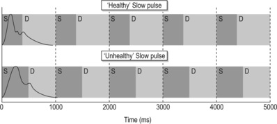 |
| Figure 6.2Variation between the systolic (S) and diastolic (D) components of the pulse wave between the formation of a healthy slow pulse and an unhealthy slow pulse. |
| Pulse presentation | Signs and Symptoms | Physiological response | |
|---|---|---|---|
| Yang vacuity | Slow and forceless | Desire for warmth, no thirst, bright-pale face, cold limbs | Long systole, small pulse amplitude |
| Pathogenic cold | Slow and forceful | Pain, thirst for warm drinks, pale face, cold limbs | Normal systole, increased amplitude, increased arterial tension |
| Pathogenic heat | Slow and forceful | Heat signs such as fever, abdominal pain | Normal systole, increased amplitude |
| Health | Slow and forceful | No cold or heat signs. No aversion to cold or heat | Short systole, long diastole |
There is also a third differential factor, but this is more subjective or sensory dependent. Constant (1999: p. 32) describes this as the sense of a ‘tap’ against the fingers. When the heart contracts strongly and quickly, the pulse wave also expands quickly to its peak and this is felt as a ‘tap’ against the fingers. When the heart contraction is slower, the pulse may rise or lift against the finger but there is no tap.
Slow pulse indicating pathology
According to CM theory, ‘When Qi moves, blood moves’ (Flaws 1994: p. 58). Therefore decreased movement of Qi leads to decreased flow rate of blood. This occurs in two ways:
• The presence of strong pathogenic Cold may result in a decrease in pulse rate. ‘Cold leads to the contraction of Qi’ (Flaws 1994: p. 59) and this retards the movement of Qi and blood.
• A vacuity of Yang Qi may result in a pulse rate slower than normal, as there is insufficient Yang Qi to propel the blood normally.
As a pathological pulse type, the Slow pulse may indicate three possible patterns:
Invasion of external pathogenic cold (Slow and forceful)
Cold has a contracting nature, causing contraction of both blood vessels and muscles and resulting in constrained flow of Qi and blood. It acts as a counter to the Yang’s warming and expansive nature and constrains the movement of Qi, contributing to a slowing of the heart rate. The presence of an external pathogenic factor will be felt as an increase in the force of the pulsation. (See the Floating pulse and the Tight pulse for specifics on the pulse in the presence of an external pathogenic agent (EPA) of Cold).
Vacuity of Yang Qi (Slow and forceless)
Yang Qi provides the motive force to move blood. ‘When Qi moves, blood moves’ (Flaws 1994: p. 58). Consequently, deficient Yang Qi will lead to impaired flow of blood. The Nei Jing notes in Chapter 18 that, compared to that of a ‘normal’ pulse, a pulse that beats only twice per complete respiration signifies a deficiency of Qi.
When man exhales once and his vessels exhibit one movement and when he inhales once and his vessels exhibit one movement, that is called ‘short of Qi’.
Pathogenic heat
Lu (1996) describes an occurrence of the Slow pulse in the presence of pathogenic Heat in the intestines causing obstruction. Heat causes fluids to congeal, so obstruction of Yang Qi leads to a Slow pulse, but the pulse is forceful and accompanied by signs and symptoms associated with heat rather than cold signs.
6.4.1.5. Biomedical perspective
Termed bradycardia, the pathological Slow pulse is defined as having a heart rate slower than 60 bpm which is accompanied by a systole that is slower (or longer) than normal. This can be due to a number of factors including low body temperature, medication such as digitalis, hypothyroidism, heart problems and electrolyte imbalances (Ca2+, Na+, K+). Carotid sinus syndrome can result in bradycardia due to hypersensitivity of the carotid artery walls to pressure. Mild external pressure triggers a strong baroreceptor reflex which results in vagal stimulation that has a parasympathetic effect, thus slowing the heart rate (Guyton & Hall 2006: p. 148).
Pathologically, an extremely slow pulse may be due to heart block, where there is impairment of the normal electrical conduction pathway through the heart that causes normal cardiac contraction. (In CM this is viewed as Heart Yuan Qi vacuity.) There are different degrees of heart block, linked to the level of impairment of the heart’s conduction system. Signs range from delayed heart contraction through to compromised circulation and eventual heart failure. Treatment protocols also vary, ranging from medications to physical insertion of an artificial pacemaker to replace the heart’s own natural pacemaker (SA node: see section 6.3.5).
6.4.2. Rapid pulse (Shuò mài)
6.4.2.1. Requisite parameters
The Rapid pulse is a simple pulse quality defined only by changes in the pulse rate parameter. There is an increase in pulse rate above that of the normal heart rate range, 60–90 bpm.
6.4.2.2. Clinical definition
The rate of the Rapid pulse is greater than or equal to 90 bpm.
Note that the rate of 90 bpm for classifying a rapid pulse is different from the biomedical definition of a rapid pulse or tachycardia, commonly listed as 100 bpm. See 6.4.2.5 Biomedical Perspective, below, for further information. Also see Box 6.3, Racing pulse.
Box 6.3
Racing pilse 
There is a subcategory of the Rapid pulse called the Racing pulse. The Racing pulse is defined by a pulse rate greater than 120 bpm, or seven or more beats per respiration cycle. According to Deng (1999: p. 113), the pulse was first described in the Zhen Jia Shu Yao around 1000 ce, and expanded on in the Ming dynasty text Zhen Jia Zheng Yan . The Racing pulse is similar to the biomedical definition of tachycardia, with a similar description of aetiology and conditions in which it manifests such as thyrotoxicosis and high fevers reflecting an increased metabolic rate. It may be associated with some forms of compromised cardiac function.
6.4.2.3. Classical description from the Mai Jing and Nei Jing
The rapid pulse is a pulse coming and going abruptly and urgently [beating 6–7 times in one respiration in another version; named an advancing (pulse) in yet another]
(Mai Jing; (Wang, Yang (trans) 1997: p. 3).
Rapid is defined as more than five beats per breath of the doctor
(Nei Jing;Ni (trans) 1995: p. 31).
6.4.2.4. CM indications for the Rapid pulse
The Rapid pulse may indicate health or pathology depending on the age of the individual.
Health
In children, a rapid pulse is considered ‘auspicious’ or favourable (Li, Flaws (trans) 1998: p. 75), as children’s pulses are usually more rapid than those of adults. This is attributable to their Yang nature and so a rapid pulse is an appropriate pulse to manifest. The Mai Jing notes specifically that ‘In children between four and five years, the pulse is fast, beating 8 times per respiration’ (Wang, Yang (trans) 1997: p. 10). The normal pulse rate for adults is described as 4–5 beats per respiration, which means that the normal children’s pulse rate is twice that of adults when using the old respiration method.
Pathology
In an adult, the Rapid pulse always indicates pathology involving heat or hyperactivity of Yang. Any hyperactivity of Yang Qi may augment pulse movement of Qi and blood, resulting in an increased heart rate. This may indicate the presence of pathogenic Heat or Fire, as seen in febrile diseases. Alternatively, a rapid pulse may also result from an inability of Yin to control Yang, allowing Yang to move without its usual constraints:
When the Yin fails to contain the Yang, the flow in the channels will become rapid, causing the Yang Qi to become excessive and reckless.
(Wei Jing Su Wen)
This may be due to a deficiency of Yin fluids such as blood or body fluids arising from blood loss in haemorrhage, or depletion of Yin fluids following febrile disease, through excessive sweating or diarrhoea. Accordingly, two main patterns are associated with a pathological Rapid pulse:
An increase in Yang supplements the body’s Yang Qi in moving Qi and blood. Consequently, the pulse rate increases (changes in other parameters such as contour and possibly arterial width and length may also be present). Repletion of Yang is often attributable to external variables, such as the presence of an external pathogenic factor producing fever. Dietary factors such as alcohol, herbs and spices may also supplement the body’s Yang. In this situation the pulse will be Rapid and forceful (see section 6.8.1 for further information).
Yin vacuity producing heat (rapid and forceless)
Yin vacuity may occur as the result of loss of Yin fluids through excessive sweating, vomiting, diarrhoea or blood loss. As the relative amount of Yin (in the form of fluids) has decreased then there is a relatively excessive amount of Yang producing heat. As such, the term, ‘Yin vacuity producing heat’ refers to the creation of heat in the body because Yin is vacuous. Poor sleep and overwork are lifestyle factors which have an cumulative affect over time of depleting Yin.
From a biomedical perspective Yin vacuity can be associated with a relative increase in the sympathetic nervous system due to an inability of the parasympathetic nervous system to maintain appropriate control over these aspects. For example, stress can cause palpitations (see section 6.5.4) and digestive disturbances (Wood attacking the Earth); conditions such as irritable bowel syndrome (IBS) are affected by stress, producing bouts of either constipation or diarrhoea. Additionally, Guyton & Hall (2000) note that the ‘contractile strength of the heart often is enhanced temporarily by a moderate increase in temperature, but prolonged elevation of the temperature exhausts the metabolic systems of the heart and eventually causes weakness’ (p. 106).
There are two possible effects on the pulse wave when Yin becomes vacuous. These are:
– If Yin is vacuous then it is no longer able to control (or constrain) the movement and function of Yang. Therefore the flow rate of Qi and Blood in the arteries increases, manifesting as an increase in heart rate.
– As Yin’s function of anchoring Yang may be affected, the pulse could also present relatively stronger (but overall is not actually forceful) at the superficial level as Yang ‘floats’, moving upwards and outwards, in addition to an increase in heart rate.
6.4.2.5. Biomedical perspective
Tachycardia or tachyarrythmia is defined as an abnormally fast heart rate (>100 bpm) that may be transient or ongoing. When heart rate increases there is inefficient filling of the left ventricle during diastole and stroke volume is diminished. Whether due to poor refill during diastole or shortened ejection duration with systole, the result of tachycardia is often a compromised circulation of blood affecting tissue perfusion, especially over the longer term (Box 6.3).
Guyton & Hall (2006: p. 147) note three general causes of tachycardia:
• Increased body temperature
• Sympathetic nervous system stimulation of the heart
• Toxic conditions of the heart.
Tachycardia may also result from acute emotional stress such as anxiety, increased body temperature associated with fever or exercise (Box 6.4), blood loss and anaemia reducing effective blood volume or medication stimulating the pacemaker, or as a reflexive response from heart disease in an attempt to maintain normal circulation. Box 6.5 lists some conditions associated with tachycardia.
Box 6.4
Heart rate and temperature
Guyton & Hall (2006: p. 197) assert that heart rate increases by 18 bpm for each °Celsius increase in body temperature to 40.5 °Celsius. Beyond this the heart weakens and so pulse rate may slow. HR increases with fever because the increased temperature stimulates the SA node’s metabolic rate, excitability rate of rhythm.
Box 6.5
Conditions associated with tachycardia
• Sinus tachycardia
• Atrial and ventricular fibrillation (heart Qi or Blood stagnation; heart shock)
• Hyperthyroidism (which may equate to Yin vacuity)
• Febrile diseases (Yang excess)
• Haemorrhage
• Pregnancy. In the third trimester normal resting heart rate increases by 10 bpm and cardiac output increases by 40% due to increased stroke volume (Braunwald et al 2001: p. 25)
• Chronic rheumatic heart disease may result in damage to the heart structure such as heart valves, as well as conduction defects leading to atrial fibrillation
From both CM and biomedical perspectives, the conditions listed above are accompanied by changes in other pulse parameters, not just pulse rate alone.
The body’s response to stress is a common cause of tachycardia. Stress initiates neuroendocrine processes, leading to the release of epinephrine (adrenaline) and norepinephrine (noradrenaline), as well as other hormones from the pituitary and adrenal glands (‘fright, flight or fight’ response). Norepinephrine (noradrenaline) constricts smooth muscle in all blood vessels and therefore plays a major role in peripheral vasoconstriction. Epinephrine (adrenaline) is responsible for increasing heart rate and also for the force of myocardial contraction.
Atrial fibrillation is a form of tachycardia, involving increased impulses affecting the contraction of the atria. (A number of the CM pulse qualities are due to atrial fibrillation.) As atrial fibrillation can also result in irregular rhythm, it is discussed in section 6.5 on Rhythm.
Hyperthyroidism increases heart rate considerably because the thyroid hormone appears to directly cause heart excitation. Thyroid hormone may also increase cardiac output and blood flow (Guyton & Hall 2006: p. 937).
Haemorrhage may also cause an increase in heart rate, as part of a cardiovascular compensatory mechanism to increase circulating volume by pulling fluids from the interstitium. This dilutes the viscosity of blood, resulting in faster blood flow. In addition, hypoxia results in vasodilatation of blood vessels, also increasing blood flow. As a result venous blood return is increased and the heart needs to pump faster and harder to get oxygenated blood to the body and prevent cardiopulmonary congestion (McCance & Huether 2006: p. 929).
6.4.3. Moderate pulse (Hûan mài) 
6.4.3.1. Alternative names
Leisurely, Slowed Down, Relaxed, Retarded or Languid pulse.
6.4.3.2. Requisite parameters
The Moderate pulse is a simple pulse that has changes in two pulse parameters:
• Rate: There may be either an increase or decrease in rate dependent on the individual’s ‘normal’ resting heart rate.
• Contour: The pulse shape is rounded (see Slippery pulse, section 7.9.1).
The Moderate pulse is initially identified by the pulse rate (Table 6.4) and further differentiated from the standard Slow pulse by the shape of the pulse contour.
| Pulse rate (bpm) | Pulse quality/category | Biomedical | Possible indications | Traditional (Beats per respiration) |
|---|---|---|---|---|
| <60 | Slow pulse | Bradycardia | Health | 3 |
| Cold EPA Heath Damp | ||||
| Heath Damp | ||||
| 60 | Moderate pulse Slow pulse | Yang vacuity | 4 | |
| 61–80 | Normal pulse rate | Normal pulse rate | Health | 4-5 |
| 81–89 | Borderline normal to rapid | Health | 5–6 | |
| Unhealthy heart | ||||
| Blood vacuity | ||||
| Recovery phase | ||||
| Progression of illness | ||||
| 90–119 | Rapid pulse | Tachycardia | EPA Heat | 7-8 |
| Vacuity Heat | ||||
| >120 | Racing pulse | Heat/Fire | ≥8 | |
| Infections | ||||
| Dehydration | ||||
| Extreme fevers |
6.4.3.3. Clinical definition
The Moderate pulse has a pulse rate of 60 bpm, with a distinctly rounded contour felt under the palpating fingers.
6.4.3.4. Classical descriptions from the Nei Jing and Mai Jing
A pulse that is neither too strong nor too weak, that comes and goes in a rhythmic fashion, flowing like a stream … (Nei Jing)
The moderate pulse is also a pulse slow in coming and going but a little faster than the slow pulse …’ (Mai Jing)
6.4.3.5. CM indications
The Moderate pulse can be either healthy pulse or pathological, depending on the signs and symptoms accompanying it.
Health
It is a normal pulse rate in a healthy person if no other change in pulse characteristics accompany it, or when there are no other presenting pathological signs and symptoms. In this situation it is a sign of a strong constitution. When this occurs, it could be classified both as a Slow pulse, due to the rate, but also a Slippery pulse, and would be a sign of sufficient Qi and blood.
Pathology
The Moderate pulse is often associated with the presence of Damp or vacuity of the Stomach and Spleen, especially Spleen Yang Qi deficiency. Therefore, it would occur in conjunction with signs and symptoms associated with such patterns. This could include digestive symptoms such as loose stools, fatigue and tiredness, cold limbs, fluid retention and an aversion to cold.
Pathogenic damp is broadly seen as a Yin condition and tends to injure Yang. In addition, damp is heavy, slowing down and impeding the flow and circulation of Qi and blood. ‘Phlegm and Dampness lead to Qi obstruction; Qi obstruction leads to Qi stagnation’ (Flaws 1994: p. 79).
6.5. Rhythm
Pulse rhythm is an expression of the heart’s functional capacity to contract and relax in a consistent fashion. Rhythm as such is not how frequently the heart contracts — this is pulse rate — rather, it is whether the heart is sufficiently contracting at all.
A normal pulse has a regular rhythm, with a consistently even interval between each pulsation. When the intervals between pulsations vary in length or there appears to be ‘missing’ beats or an interruption, this is said to be an irregular rhythm. Any pulse occurring with an irregular rhythm is termed an arrhythmia or dysrhythmia, or simply a pulse lacking a regular rhythm.
Arrhythmic pulses may have an interruption to their normal rhythm occurring at irregular or regular intervals. Interruptions range from pulses with occasional ‘missed’ beats or rapid beats, to serious rhythmic disturbances that impair the pumping action of the heart.
Three specific CM pulse qualities are associated with the rhythm parameter:
• Skipping pulse (section 6.6.1)
• Bound pulse (section 6.6.2)
• Intermittent pulse (section 6.6.3).
The Skipping pulse and Bound pulses have irregular interruptions to their normal rhythms and are further differentiated by pulse rate. The Intermittent pulse has regularly spaced interruptions to the normal rhythm and is defined only by the rhythm parameter.
6.5.1. Pulse rhythm and its assessment
The method for assessing pulse rhythm requires the pulse to be felt for an interval of at least 60 seconds. This is because irregularly interrupted pulses, with only occasional interruptions to normal heart rhythm, may not be detected in any shorter time frame. With the fingers placed on the artery, the practitioner notes the presence of pulsations and whether these are occurring at regularly spaced intervals.
When an irregularity in rhythm is detected, such as a ‘missed’ beat, the practitioner next needs to determine the nature and frequency of the interruption to normal pulse rhythm:
• Is the interruption to heart rhythm occuring regularly (that is, is it occurring at a consistent interval between each beat?)
• If so, how often does this occur? (number of rhythmic beats between each interruption to heart rhythm)
• Or, is the interruption to heart rhythm occurring only at irregularly spaced intervals: is there a missed beat only occasionally (no regular interval or specific number of beats between each missed beat)?
When assessing pulse rhythm, it is also important to inquire if the patient is aware of any irregularity in their heart rate, as this may not be apparent at the time of consultation (Box 6.6). Changes in heart rhythm may include palpitations and this will be discussed in 6.5.4.
Box 6.6
Clinical questions to ask your patient
• Are you aware of what your normal heart rate is?
• Are you aware that it is slower or faster than usual?
• If you have noticed changes to your HR does this occur suddenly or gradually?
• How long has this been happening?
• Is this happening all the time?
• Does this occur with any other symptoms?
• Is it better when you rest?
Sometimes an interruption to the pulse rhythm can be due to a blockage in the conduction of the pulse wave from the heart to the periphery (Box 6.7). It is therefore also important to compare the left and right radial pulses. Differences in rhythm between the two sides may indicate some type of problem with the arterial system, such as arterial blockage or aortic coarctation, rather than specific heart-related pathology (Constant 1999).
Box 6.7
Sinus arrhythmia
• Sinus arrhythmia is a normal occurrence often seen in young adults where the heart rate slightly speeds up during inspiration due to activation of neural input to the brain when the lungs are expanded (for example, deep breathing), and then slows down during expiration.
• Sinus arrhythmia often results from alteration of the strength of the nerve signal to the heart sinus node affecting the heart rate (Guyton & Hall 2000: pp. 134-135). Specifically, the mechanism affecting heart rate occurs during deep breathing when neural receptors in the lungs are activated.
•McCance & Huether (2006) state that the increase in heart rate during inspiration is caused by the stretching (activation) of vagal fibres in the lungs that cause heart rate to speed up by inhibiting the cardioinhibitory centre of the medulla. Inhibition of this centre allows unopposed sympathetic acceleration of heart rate (p. 1049).
• Sinus arrhythmia needs to be carefully differentiated from arrhythmia or dysrhythmia. A pulse is described as arrhythmic when there are pauses in the heart’s normal conduction system resulting in a perceived missed beat. With sinus arrhythmia there are no missed beats but rather a change in the length of time between beats. This is not deemed a pathological occurrence.
• The phenomenon of sinus arrhythmia was likely clinically observed by the authors of the Nei Jing, as evidenced by the following lines of text.
If the vessels exhibit five movements,
this is an intercalation [of a fifth movement] because of a deep breathing.
That is called a ‘normal person’.
(Su Wen)
This partial description of the normal pulse appears be an early reference to sinus arrhythmia and was clearly viewed as a variation of the normal healthy pulse rate.
6.5.2. Regulation of pulse rhythm: CM perspective
In CM the Heart governs the movement of blood. Heart Qi maintains the functional ability of the heart to contract. Rhythm is affected when Heart Qi is affected. In this context, changes in pulse rhythm occur when the heart Qi is exhausted or obstructed.
Changes in heart rhythm, especially rhythm changes associated with cardiovascular damage, can be the final common pathway for a number of diseases (AtCor Medical 2006: pp. 1-31). These include:
• Hypertension
• Left ventricular hypertrophy and failure
• Diabetes mellitus
• Renal disease
• Hyperlipidaemia.
Differentiation of the cause and aetiology are further elucidated on other presenting signs and symptoms and medical history. In a pulse diagnosis context, the parameter of pulse rate is used to identify aetiological factors of heat and cold affecting heart rhythm. This includes internal aetiologies arising from vacuities of Yin and Yang, and external pathogenic factors of heat and cold.
In CM terms, the Heart also governs the Mind or Shen. Physical heart damage will, theoretically, affect the clear expression of the Shen and be associated with Blood stagnation. The relationship is seen in the incidence of depression and heart disease. Severe shock or pain, anxiety and stress also affect the Shen, which in turn can affect heart rhythm.
Stay updated, free articles. Join our Telegram channel

Full access? Get Clinical Tree


