Detection, localization, and quantification of intracardiac shunts are an integral part of the hemodynamic evaluation of patients with congenital heart disease. In most cases, an intracardiac shunt is suspected on the basis of the clinical evaluation of the patient before catheterization. There are several circumstances, however, in which data obtained at catheterization should alert the cardiologist to look for a shunt that had not been suspected previously:
1. Unexplained arterial desaturation should immediately raise the suspicion of a right-to-left intracardiac shunt, which may then be assessed by the methods to be discussed. Most commonly, arterial desaturation (i.e., arterial blood oxygen saturation <95%) detected at the time of cardiac catheterization represents alveolar hypoventilation. The causes for this alveolar hypoventilation and its associated physiologic right-to-left shunt include (a) excessive sedation from the premedication, (b) chronic obstructive lung disease or other pulmonary parenchymal disease, and (c) pulmonary congestion/edema secondary to the patient’s cardiac disease. Alveolar hypoventilation associated with each of these problems is exacerbated by the supine position of the patient during the catheterization procedure. Helping the patient to assume a more upright posture (head-up tilt or propping the patient up with a large wedge if tilt mechanism is not available) and encouraging the patient to take deep breaths and to cough will correct or substantially ameliorate arterial hypoxemia in most cases. If arterial desaturation persists, oxygen should be administered by face mask for both therapeutic and diagnostic purposes. If full arterial blood oxygen saturation cannot be achieved by face-mask administration of oxygen (it is best in this regard to use a rebreathing mask that fits snugly), a right-to-left shunt is presumed to be present, and its anatomic site and magnitude should be determined using the methods described later in this chapter.
2. Conversely, when the oxygen content of blood in the pulmonary artery is unexpectedly high (i.e., if the pulmonary artery [PA] blood oxygen saturation is >80%), the possibility of a left-to-right intracardiac shunt should be considered. It is for these two reasons that arterial and pulmonary artery saturation should be measured routinely during cardiac catheterization.
3. When the data obtained at cardiac catheterization do not confirm the presence of a suspected lesion, one should consider the presence of an intracardiac shunt. For example, if left ventricular cineangiography fails to reveal mitral regurgitation in a patient in whom this was judged to be the cause of a systolic murmur, it is prudent to look for evidence of a ventricular septal defect (VSD) with left-to-right shunting.
DETECTION OF LEFT-TO-RIGHT INTRACARDIAC SHUNTS
Many techniques are available for the detection, localization, and quantification of left-to-right intracardiac shunts. The techniques vary in their sensitivity, the type of indicator they use, and the equipment needed to sense and read out the presence of the indicator.
Measurement of Blood Oxygen Saturation and Content in the Right Heart (Oximetry Run)
In the oximetry run, a basic technique for detecting and quantifying left-to-right shunts, the oxygen content or percentage saturation is measured in blood samples drawn sequentially from the pulmonary artery, right ventricle (RV), right atrium (RA), superior vena cava (SVC), and inferior vena cava (IVC). A left-to-right shunt may be detected and localized if a significant step-up in blood oxygen saturation or content is found in one of the right heart chambers. A significant step-up is defined as an increase in blood oxygen content or saturation that exceeds the normal variability that might be observed if multiple samples were drawn from that cardiac chamber.
The technique of the oximetry run is based on the pioneering studies of Dexter and his associates in 1947.1 They found that multiple samples drawn from the right atrium could vary in oxygen content by as much as 2 volumes percent (vol%).a This variability has been attributed to the fact that the right atrium receives its blood from three sources with varying oxygen content: the superior vena cava, inferior vena cava, and coronary sinus. The maximal normal variation within the right ventricle was found to be 1 vol%. Because of more adequate mixing, a maximal variation within the pulmonary artery of only 0.5 vol% was found by Dexter. Thus, using the Dexter criteria, a significant step-up is present at the atrial level when the highest oxygen content in blood samples drawn from the right atrium exceeds the highest content in the venae cavae by 2 vol%. Similarly, a significant step-up at the ventricular level is present if the highest oxygen content in the right ventricular samples is 1 vol% higher than that in the highest right atrial sample, and a significant step-up at the level of the pulmonary artery is present if the pulmonary artery oxygen content is more than 0.5 vol% higher than the highest right ventricular sample.
Dexter’s study described normal variability and gave the criteria for a significant oxygen step-up only for measurement of blood oxygen content. This in part reflects the methodology available to him, because spectrophotometric oximetry was not widely used at that time. In recent years, nearly all cardiac catheterization laboratories (especially those primarily involved in pediatric catheterization) have moved toward the measurement of percentage oxygen saturation by spectrophotometric oximetry as the routine method for oximetric analysis of blood samples. Oxygen content may then be calculated from knowledge of percentage saturation, the patient’s blood hemoglobin concentration, and an assumed constant value for the oxygen-carrying capacity of hemoglobin (1.36 mL O2/g hemoglobin), as discussed in Chapter 11. When oxygen content is derived in this manner, rather than by measurement by the Van Slyke or other direct oximetric technique, the value is no more accurate (and probably less so because of the potential presence of carboxyhemoglobin or hemoglobin variants with O2-carrying capacity other than 1.36) than the percentage oxygen saturation values from which it is calculated.
To clarify this situation, Antman and coworkers prospectively studied the normal variation of both oxygen content and oxygen saturation of blood in the right heart chambers.2 The study population consisted of patients without intracardiac shunts who were undergoing diagnostic cardiac catheterization for evaluation of coronary artery disease, valvular heart disease, cardiomyopathy, or possible pulmonary embolism. Each patient had a complete right heart oximetry run (see later discussion) with sampling of multiple sites in each chamber. Oxygen content was measured directly by an electrochemical fuel cell method (Lex-O2-Con, Lexington Instruments, Lexington, MA), a method that had been previously validated against the Van Slyke method. Oxygen saturation was calculated as blood oxygen content divided by oxygencarrying capacity. The relationship between oxygen content and oxygen saturation obviously depends on the hemoglobin concentration in the patient’s blood (e.g., 75% oxygen saturation of pulmonary artery blood will be associated with a substantially lower oxygen content in an anemic patient than in one with normal hemoglobin concentration). Also, systemic blood flow may be an important determinant of oxygen variability in the right heart chambers because high systemic flow tends to equalize the differences across various tissue beds.
In the context of these considerations, listed in Table 12.1 are the criteria for a significant step-up in right heart oxygen content and percentage oxygen saturation associated with various types of left-to-right shunt, based on the study of Antman and coworkers2 and other investigators.1,3,4,5,6 As can be seen from the bottom line (“Any level”) of Table 12.1, the simplest way to screen for a left-to-right shunt is to sample SVC and PA blood and measure the difference, if any, in percentage O2 saturation. It is recommended that blood samples from SVC and PA be routinely obtained at the time of right heart catheterization and their O2 saturation determined by reflectance oximetry. If the ΔO2 saturation between these samples is ≥8%, a left-to-right shunt may be present at atrial, ventricular, or great vessel level, and a full oximetry run should be done.
Oximetry Run
The blood samples needed to localize a step-up in the right heart are obtained by performing what is called an oximetry run. The samples needed and the recommended order in which they should be obtained follow.
Obtain a 2 mL sample from each of the following locations:
1. Left and/or right pulmonary artery
2. Main pulmonary arteryb
3. Right ventricle, outflow tractb
4. Right ventricle, midc
5. Right ventricle, tricuspid valve or apexbc
6. Right atrium, low or near tricuspid valve
7. Right atrium, mid
8. Right atrium, high
Table 12.1 Detection of Left-to-Right Shunt by Oximetry
SVC and IVC, superior and inferior vena cavae; RA, right atrium; RV, right ventricle; PA, pulmonary artery; VSD, ventricular septal defect; TR, tricuspid regurgitation; PDA, patent ductus arteriosus; PR, pulmonic regurgitation; ASD, atrial septal defect; SBFI, systemic blood flow index; Qp/Qs, pulmonary to systemic flow ratio.
9. Superior vena cava, low (near junction with right atrium)
10. Superior vena cava, high (near junction with innominate vein)
11. Inferior vena cava, high (just at or below diaphragm)
12. Inferior vena cava, low (at L4-L5)
13. Left ventricle
14. Aorta (distal to insertion of ductus)
In performing the oximetry run, an end-hole catheter (e.g., Swan-Ganz balloon flotation catheter) or one with side holes close to its tip (e.g., a Goodale-Lubin catheter) is positioned in the right or left pulmonary artery. Cardiac output is measured by the Fick method. As soon as the determination of oxygen consumption is completed, the operator begins to obtain 2 mL blood samples from each of the locations indicated. This is done under fluoroscopic control, with catheter tip position further confirmed by pressure measurement at the sites noted. The entire procedure should take less than 7 minutes. If a sample cannot be obtained from a specific site because of ventricular premature beats, that site should be skipped until the rest of the run has been completed.
Oxygen saturation and/or content in each of the samples is determined as discussed previously, and the presence and localization of a significant step-up are determined by applying the criteria listed in Table 12.1.
An alternative method for performing the oximetry run is to withdraw a fiberoptic catheter from the pulmonary artery through the right heart chambers and the inferior and superior venae cavae. This permits a continuous readout of oxygen saturation that allows detection of a step-up in oxygen content.
If the oximetry run reveals that a significant step-up is present, the pulmonary blood flow, systemic blood flow, and magnitude of left-to-right and right-to-left shunts may be calculated according to the following formulas.
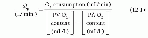
Calculation of Pulmonary Blood Flow (Qp)
Pulmonary blood flow is calculated by the same formula used in the standard Fick equation:
If a pulmonary vein (PV) has not been entered, systemic arterial oxygen content may be used in the preceding formula, if systemic arterial oxygen saturation is ≥95%. If systemic oxygen saturation is <95%, one must determine whether a right-to-left intracardiac shunt is present. If there is an intracardiac right-to-left shunt, an assumed value of 98% oxygen capacity for the pulmonary venous oxygen content should be used in calculating pulmonary blood flow. If arterial desaturation is present and is not owing to a right-to-left intracardiac shunt, the observed systemic arterial oxygen saturation should be used to calculate pulmonary blood flow.
Example
Let us suppose that a patient is found to have an atrial septal defect (ASD) with a left-to-right shunt clearly detected by oximetry run. Furthermore, the catheter crosses the defect and a pulmonary vein is entered, from which a blood sample shows O2 saturation of 98%. Let us further suppose that systemic arterial blood saturation, however, is 90% and that this is owing to chronic pulmonary disease. After ruling out a right-to-left shunt by any of the various methods (e.g., inhalation of 100% oxygen, indocyanine green dye injection in inferior vena cava, echocardiogram-bubble study), should we use 98% or 90% for pulmonary venous blood O2 saturation in the calculation of Qp? As indicated earlier, because arterial desaturation is not caused by a right-to-left intracardiac shunt, the observed systemic arterial O2 saturation (90%) should be used because this summates all the pulmonary veins draining both lungs, not just the one with 98% O2 saturation.
Table 12.2 Calculation of Systemic Blood Flow in the Presence of Left-to-Right Shunt
Location of Shunt as Determined by Site of O2 Step-Up
Mixed Venous Sample to be Used in Calculating Systemic Blood Flow
Right ventricle, average of samples obtained during oximetry run
2. Right ventricle (e.g., ventricular septal defect)
Right atrium, average of all samples during oximetry run
3. Right atrium (e.g., atrial septal defect)
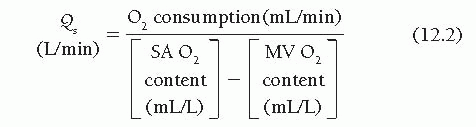
Calculation of Systemic Blood Flow (Qs)
Use the following equation for systemic blood flow:
The key to proper measurement of systemic blood flow in the presence of an intracardiac shunt is that the mixed venous oxygen content must be measured in the chamber immediately proximal to the shunt, as shown in Table 12.2.
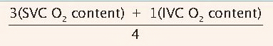
The formula generally used by cardiologists who treat adults for the calculation of venous content in the presence of an ASD was derived by Flamm and coworkers.5 They found that systemic blood flow calculated from mixed venous oxygen content as determined from the formula listed in Table 12.2 most closely approximates systemic blood flow as measured by left ventricular to brachial artery (BA) dye curves in patients with atrial septal defect studied at rest. It should be noted that Flamm’s formula weights blood returning from the superior vena cava more heavily than might be expected on the basis of relative flows in the superior and inferior cavae. The success of this empirical weighting of the relatively desaturated superior vena cava blood (O2 saturation is almost always less in blood from the superior as opposed to the inferior vena cava) probably reflects the fact that the third contributor to mixed venous blood—desaturated coronary sinus blood—is not sampled during the oximetry run and therefore cannot be included directly in the formula. The formula (3 SVC O2 + 1 IVC O2)/4 was validated by Flamm and associates for mixed venous oxygen content at rest5: In 18 patients without shunt, this value agreed closely with pulmonary artery blood oxygen content at rest. During supine bicycle exercise, however, a different relationship was found to apply, in which mixed venous (pulmonary artery) oxygen content in patients without shunt was best approximated as (1 SVC O2 + 2 IVC O2)/3. This formula was then used for patients with atrial septal defect during exercise, and it reliably predicted systemic blood flow measured by left ventricular to BA dye-dilution curve. Therefore, for patients with left-to-right shunt at the atrial level, the formula in Table 12.2 should be used only for calculation of resting mixed venous O2 content.
Only gold members can continue reading. Log In or Register to continue