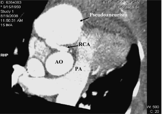
Magnetic resonance imaging (MRI) revealed a large rounded mass between the myocardium and pericardium lying in the lateral aspect of the right atrioventricular groove with flow into the mass (Videoclip 18.2). There was significant pericardial scarring and inferior left ventricle (LV) hypokinesis. Computed tomography (CT) angiogram revealed a mid right coronary artery (RCA) aneurysm that appeared to have ruptured distally causing a large pseudoaneurysm (Figure 18.2) and a fistula between the pseudoaneurysm and right ventricle (RV) through which the pseudoaneurysm was emptying its contents during diastole (Videoclip 18.3). The proximal and distal portions of the RCA were noted to be heavily calcified.
Figure 18.2 This large pseudoaneurysm is from the right coronary artery and is well demonstrated by CT image. AO, aorta; PA, pulmonary artery; RCA, right coronary artery.

Stay updated, free articles. Join our Telegram channel

Full access? Get Clinical Tree


