Role of Transesophageal Echocardiography in Mitral Valve Repair

20 Role of Transesophageal Echocardiography in Mitral Valve Repair


Mitral Valve Diagrams


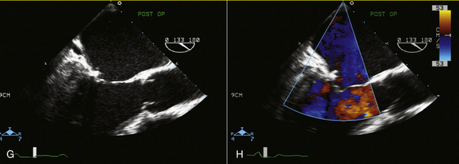
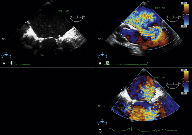
Figures 20-1 through 20-5 present the anatomy of the mitral valve.


image

Figure 20-3 This diagram was designed in 1995 by the Toronto General Hospital Echocardiography Laboratory to identify the anatomopathology of the mitral valve by transesophageal echocardiography (TEE) to predict suitability for repair.1 In this diagram, the mitral valve is viewed as if looking from the apex to the base of the heart. The transesophageal probe is positioned in the esophagus and is directed toward the mitral valve from posterior to anterior. Starting at the 9 o’clock position, to the left of the diagram, the corresponding TEE picture is shown. At 0 degrees, the ultrasound beam from right to left is crossing the mitral valve; therefore, P2 and A2 are visualized on this view as corresponding with the four-chamber TEE view. At 45 degrees, the ultrasound beam is cutting across the mitral valve from right to left. The beam crosses the large P2 scallop and will then cut through A2 and A3 in the anterior leaflet. At 75 degrees, the ultrasound beam crosses the left atrial appendage (LAA) from right to left, then the small P1 scallop, then, in the middle, P2 and A2 together, and finally, P3 of the posterior leaflet. This view also is called a commissural view. The LAA should be seen at the right side of the image and is the landmark for this view. At 105 degrees, the ultrasound beam, from anterior to posterior, crosses A1 first, followed by A2 and P2. At 135 degrees, which is the long axis of this view of the heart, the ultrasound beam crosses the aortic root and then A2 and P2. A1, anterior segment; A2, middle segment; A3, posterior segment; Ao, aorta; P1, anterolateral scallop; P2, middle scallop; P3, posteromedial scallop.


(From Omran AS, Woo A, David TE, et al. Transesophageal echocardiography accurately predicts mitral valve anatomy and suitability for repair. J Am Soc Echocardiogr. 2002;15:950–957. Used with permission.)


image

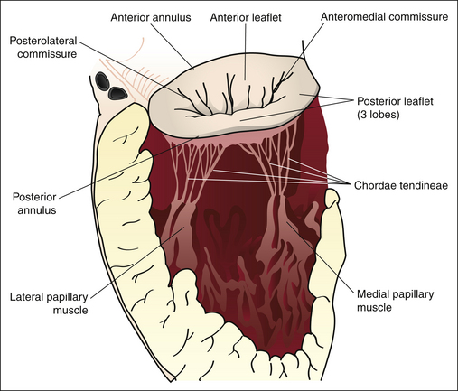
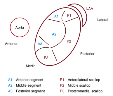
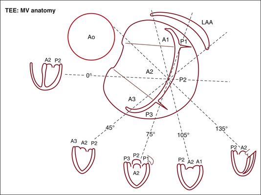
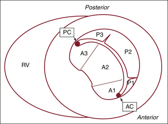
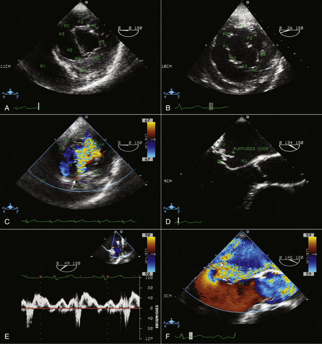
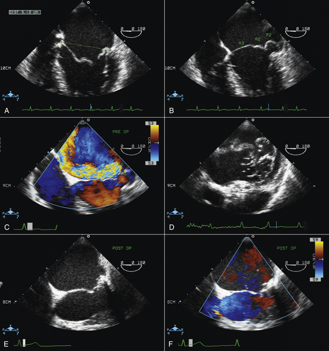
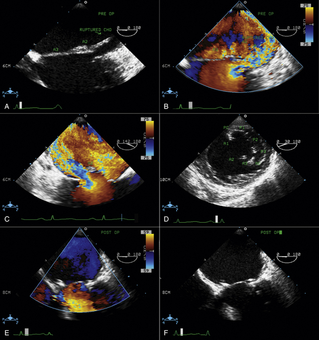
Figure 20-4 Transgastric short-axis view of the mitral valve at the level of the base of the left ventricle. This diagram was designed in the Toronto General Hospital Echocardiography Laboratory using the same classification as the illustration in Figure 20-3. Note the anterior (anterolateral) commissure (AC) at the bottom of the illustration and the posterior (posteromedial) commissure (PC) at the top. On this view, the site of origin of the mitral regurgitation can be defined by color Doppler flow mapping. A1, anterior segment; A2, middle segment; A3, posterior segment; P1, anterolateral scallop; P2, middle scallop; P3, posteromedial scallop; RV, right ventricle.


image

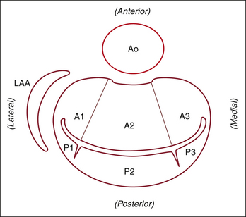
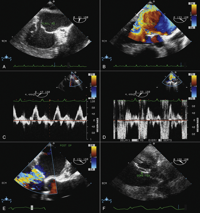
Figure 20-5 The same view as Figure 20-4, rotated 90 degrees to mimic the surgical view of the mitral valve looking from the left atrium toward the left ventricle. A1, anterior segment; A2, middle segment; A3, posterior segment; Ao, aorta; LAA, left atrial appendage; P1, anterolateral scallop; P2, middle scallop; P3, posteromedial scallop.


image image

Figure 20-6 A, Nomenclature of mitral valve anatomy in the transgastric short-axis view. A1, anterior leaflet; lateral segment; A2, anterior leaflet, middle segment; A3, anterior leaflet, medial segment; AC, anterior commissure; LV, left ventricle; P1, posterior leaflet; lateral scallop; P2, posterior leaflet; middle scallop; P3, posterior leaflet; medial scallop; PC, posterior commissure; RV, right ventricle. B, Transgastric short-axis view of the mitral valve showing a flail P2 segment. C, Transgastric short-axis view of the mitral valve showing severe mitral regurgitation due to flail P2. The regurgitant jet is originating from the site marked by the arrow. D, Midesophageal transesophageal echocardiography (TEE) provides a long-axis view of the mitral valve showing flail P2 with a ruptured chorda tendina. Mild prolapse of the middle segment (A2) of the anterior leaflet can be seen. E, Pulsed-wave Doppler interrogation of the left upper pulmonary vein shows late systolic reversal flow, compatible with severe MR, as also demonstrated by color Doppler flow mapping (F). G, Postoperative assessment of mitral valve repair showing good coaptation of the mitral leaflets. The posterior annuloplasty ring can be appreciated. H, Color flow assessment of the mitral valve after repair shows no residual mitral regurgitation. Doppler assessment of mitral inflow after mitral valve repair revealed a mean gradient of 2 mm Hg, which is normal. Note that in the postoperative assessment of mitral inflow gradient, the patient usually is tachycardic, and it is better to measure the gradient following a premature ventricular contraction.


Case 20-2


A 38-year-old man with a history of pulmonary edema.


image

Figure 20-7 A, Preoperative transesophageal echocardiography (TEE). A four-chamber view of the mitral valve is obtained to measure mitral annular (MA) dilation at end-diastole. MA = 5.1 cm, which is categorized as severely dilated. B, Four-chamber view of the mitral valve showing severe prolapse of P2 and mild prolapse of A2 and A3. The mitral annular disjunction of the posterior annulus measured 1 cm, consistent with advanced Barlow’s disease, which makes mitral valve repair technically more demanding. A2, middle segment; A3, posterior segment; P2, middle scallop. C, Four-chamber color-flow view of the mitral valve, depicting a severe anteriorly directed jet of mitral regurgitation consistent with more severe involvement of the posterior leaflet. No central or posteriorly directed jet of MR is seen, indicating that prolapse of the anterior mitral valve leaflet (AMVL) is not creating any mitral regurgitation. D, Low esophageal TEE view of the mitral valve shows severe bulky mitral leaflets with increased aneurysmal surface area of the leaflets, which is seen in very advanced Barlow’s disease. E, Postoperative TEE assessment of the mitral valve in systole after repair, illustrating very nice coaptation. This mitral valve had a complex repair with quadrangular resection of P2 and two Gore-tex loops to P1. A posterior annuloplasty ring was inserted. Preoperative prolapse of A2 was due to lack of apposition and was resolved without the need for any intervention. F, Postoperative assessment of the mitral valve with color flow Doppler showing no residual mitral regurgitation.


Jun 12, 2016 | Posted by in CARDIOLOGY | Comments Off on Role of Transesophageal Echocardiography in Mitral Valve Repair

Full access? Get Clinical Tree

Get Clinical Tree app for offline access