Right Atrial Enlargement
Gregory Kicska, MD, PhD
DIFFERENTIAL DIAGNOSIS
Common
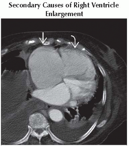
Secondary Causes of Right Ventricle Enlargement
Tricuspid Valve Disease
Chronic Atrial Fibrillation
Less Common
Left to Right Shunt
Rare but Important
Right Atrial Mass
Ebstein Anomaly
ESSENTIAL INFORMATION
Key Differential Diagnosis Issues
Radiograph shows rightward displacement of right-lower heart contour
End diastolic volume (maximum volume) > 90 mL/m2 highly specific for enlargement
Helpful Clues for Common Diagnoses
Secondary Causes of Right Ventricle Enlargement
Pulmonary hypertension suggested by aorta:PA ratio < 1:1 or main PA > 2.9 cm
Failure suggested by coexistent coronary artery disease
Tricuspid Valve Disease
Regurgitation commonly due to myxomatous degeneration, rheumatic heart disease in older population
Increased serotonin levels in carcinoid syndrome can generate fibrous tricuspid leaflet plaques that cause regurgitation
Chronic Atrial Fibrillation
Diagnosis suggested by ECG abnormality
Right atrial appendage should be examined for thrombus
Helpful Clues for Less Common Diagnoses
Left to Right Shunt
MR PA: aorta flow ratio > 1
Atrial septal defect (ASD)
MR bright-blood cine likely to show flow jet except in large ASD
Ventricular septal defect (VSD)
Most common left to right shunt but often not hemodynamically significant or spontaneously closes by adulthood
Partial anomalous pulmonary venous return
Most commonly from right upper lobe to SVC seen best on CT or MRA
Helpful Clues for Rare Diagnoses
Right Atrial Mass
Myxoma: Soft, pliable mass, connected to interatrial septum by thin stalk
Most often intermediate low T1-weighted signal, high T2-weighted signal
Ebstein Anomaly
Apical displacement of septal and posterior tricuspid leaflets with atrialization of proximal right ventricle
Coexistent tricuspid regurgitation and stenosis common
Image Gallery
 Axial enhanced CT shows RA
 and RV and RV  enlargement due to primary pulmonary hypertension and right heart failure. Left heart function is normal. On this admission, patient presented with atrial fibrillation. enlargement due to primary pulmonary hypertension and right heart failure. Left heart function is normal. On this admission, patient presented with atrial fibrillation.Stay updated, free articles. Join our Telegram channel
Full access? Get Clinical Tree
 Get Clinical Tree app for offline access

|