Reversal of Cerebral Blood Flow to Prevent Stroke during Percutaneous Carotid Artery Angioplasty and Stenting


Reversal of Cerebral Blood Flow to Prevent Stroke during Percutaneous Carotid Artery Angioplasty and Stenting


C. Schönholz, F.E. Parodi and J.C. Parodi


Embolization to the brain is a universal occurrence during carotid artery stenting (CAS) procedures. This has been documented by several well conducted ex vivo studies, by the routine use of transcranial Doppler monitoring, and by analyzing the blood aspirated after the use of distal occlusion balloons. It is also widely agreed upon that the goal of cerebral protection during CAS is to suppress or decrease the number of particles and/or air bubbles reaching the brain during the procedure.


Embolization occurs during every stage of CAS, but embolic protection starts once the cerebral protection device is deployed and finishes when it is retrieved; this means that embolization can still occur during the initial placement of the catheter in the aortic arch or carotid artery and can also be seen after the procedure is completed. Careful technique and limitation of instrumentation is crucial to reduce or suppress embolization during the initial phase of the procedure. Administration of antiplatelet therapy and appropriate use of stents of adequate size and cell configuration will sufficiently address most of the postprocedure embolic events. Crossing the lesion produces significant embolization (40,000 particles on average). In our study of carotid plaques after endarterectomy, we found that 24% of plaques have thrombus lining. In a study performed at Stanford, the incidence was 49%.


Impact of Embolization


Outcomes after CAS are related to multiple factors, including age of the patient, presence of symptoms, plaque morphology, cerebral functional reserve, aortic arch anatomy, experience of the operator, and type of cerebral protection device used, among others. There is no proof yet that microembolic signals or high-intensity transitory signals on transcranial Doppler studies are related to new cerebral lesions using diffusion-weighted (DW) magnetic resonance imaging (MRI) studies. Correlation between new lesions detected by DW-MRI and cognitive function is still ongoing. It is recognized, however, that high-intensity transitory signals are better tolerated in young patients with good cerebral functional reserve than in older patients with low functional reserve. It seems reasonable to say that high-intensity transitory signals cannot do any good to the brain, and their occurrence should be at least a reason for concern. Until we have conclusive evidence about the occurrence of high-intensity transitory signals during CAS and its potential damage to the brain, we would prefer to try to suppress or minimize its occurrence.


Flow reversal appears to be closer to the ideal device when CAS is undertaken. What is already known is that silent cerebral infarcts are relevant because they produce a steeper decline in cognitive function and increase the incidence of dementia. Deleterious effects of iodinated contrast media injected in the cerebral vasculature are well described. Using flow reversal, contrast can be readily aspirated once the injection has delineated the artery or arteries interrogated. Bubbles that are trapped in the delivery system were detected during stent deployment; aspiration will bring those bubbles out of the body using flow reversal.


Principles of Flow Reversal


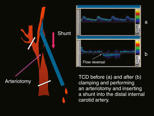
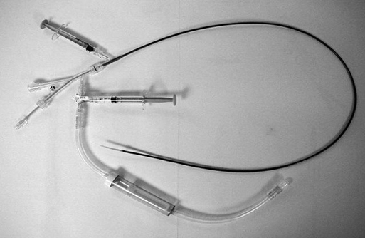
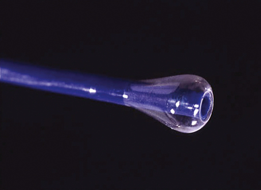
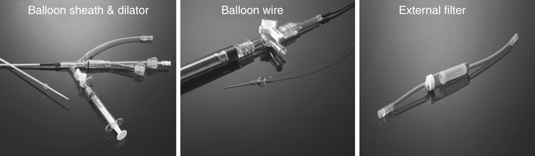
Years ago one of us (JCP) observed, during an open carotid endarterectomy using transcranial Doppler monitoring, that clamping the common carotid artery (CCA) and external carotid artery (ECA) and inserting a shunt in the distal end of the arteriotomy induced flow reversal in the middle carotid artery (MCA) if the other end of the shunt was left open to the air (Figure 1). Initially we were using the same principle through a small incision made at the base of the neck. We used a short 7-Fr introducer sheath placed in the CCA facing the lesion and occluding the ECA with a coronary balloon. The side port of the arterial line was then connected to a side port of a sheath placed in the internal jugular vein. Very soon we developed a long arterial sheath for femoral access with an occlusion balloon at its distal end (Figure 2). We designed the tip of the catheter in such a way that particles could not be trapped around the distal guiding catheter (Figure 3). We also developed an ECA occlusion balloon mounted in a hypotube and an external blood filter to be placed in between the side port of the arterial sheath and a venous sheath placed in the femoral vein.


A gradient between the internal carotid artery (ICA) and the femoral vein produces flow reversal in the ICA via collateral vessels in the circle of Willis. Similarly, intermittent aspiration enhances flow reversal during critical stages of CAS and before injecting contrast media in the guiding sheath.


This early flow reversal device became known as the Parodi AntiEmboli System (PAES) (ArteriA Medical Science, San Francisco, CA) (Figure 4). The PAES is a closed system that allows arrest of ICA flow, continuous passive ICA flow reversal, or augmented active ICA flow reversal so that any particles released during CAS pass retrograde through the catheter and are retrieved in the arteriovenous conduit filter outside the body.


The three components of the device were designed specifically to allow retrograde flow in the ICA and minimize margination of particles or collection of material that could subsequently embolize. The first component, the Parodi antiembolic catheter (PAEC), is a 9.5-Fr, 90-cm-long guide catheter with a funnel-shaped balloon on its tip. This atraumatic balloon allows occlusion of the CCA and flow reversal. It also serves as the access port for the stent delivery system and other therapeutic devices. The second component, the Parodi external balloon (PEB), is a soft atraumatic oval balloon mounted on a 0.019-inch hypotube, which is a low-profile hollow guidewire that allows inflation of the balloon. The distal shapeable, floppy guidewire facilitates navigation into the ECA. The third component, the Parodi blood return system (PBRS) is a conduit that connects the side flow reversal port of the PAEC to a venous sheath. The PBRS has a 180-μm filter that collects particulate debris before the blood reenters the venous system.

Stay updated, free articles. Join our Telegram channel

Aug 25, 2016 | Posted by in CARDIOLOGY | Comments Off on Reversal of Cerebral Blood Flow to Prevent Stroke during Percutaneous Carotid Artery Angioplasty and Stenting

Full access? Get Clinical Tree

Get Clinical Tree app for offline access