Retroperitoneal Hematoma after Angiogram
KATHERINE GALLAGHER
Presentation
A 77-year-old female presented to the post anesthesia care unit (PACU) following treatment of her left lower extremity for claudication. Per the operative report, the patient had an unremarkable procedure. An 18-gauge needle was used to cannulate the left common femoral artery, and her right superficial femoral artery (SFA) was treated with percutaneous angioplasty via an up-and-over approach. On the operative note, it was noted that the wire passed without resistance. She did receive a loading dose of 300 mg of clopidogrel.
Her medical history is significant for a coronary artery disease status plus coronary stenting, hypertension, and a 50-pack-year history for smoking. The patient has been recovering reasonably well in the PACU and has no obvious evidence of groin hematoma; however, she has had several episodes of hypotension, and she has responded to 500-mL boluses of IV fluid. She reports that she is feeling somewhat light-headed and nauseated but is without any chest pain. On examination, her pulse is 130 beats per minute and her blood pressure is 90/50. She is reporting some pain in her iliac fossa region on the left, which has been requiring analgesics. Her abdomen is soft. She has palpable bilateral femoral pulses and no evidence of hematoma in the left groin. Laboratory evaluation reveals normal creatinine, leukocyte count, and coagulation parameters; however, her hematocrit is noted to be 10 points lower than her baseline.
Differential Diagnosis
Differential diagnosis for this scenario includes complications related to the procedure (percutaneous access, perforation at the site of endovascular treatment) as well as cardiac issues (e.g., MI). The leading diagnosis is retroperitoneal hematoma as the retroperitoneum can hold a significant volume of blood without any obvious signs on physical exam. Often by the time hemodynamic compromise occurs, the patient has already lost a significant volume of blood, which can result in devastating complications in many of the vascular patients with significant heart disease and little volume reserve.
Background and Workup
Background
This patient has a potential life-threatening problem, with suspicion of retroperitoneal hematoma. First and foremost is evaluation and stabilization of the patient. Diagnostic evaluation can include abdominal CTA in order to evaluate for retroperitoneal hematoma; however, prompt volume resuscitation preferably with blood/colloid is warranted. Although most retroperitoneal hematomas can be treated conservatively, extravasation may require surgical treatment or endovascular management. Patients who continue to have persistent hypotension despite adequate resuscitation may require operative intervention. Additionally, those who fail to respond appropriately following transfusion may warrant imaging in the endovascular suite.
Patients who develop femoral neuropathy require hematoma evacuation and exploration. Endovascular embolization or stent graft placement for active extravasation is occasionally necessary. What is most important in management of the retroperitoneal hematoma is prompt recognition of the complication. Delays in recognition add to increase morbidity and mortality. Mortality rates approach 0.5% to 2% in patients who develop this complication. The most common symptoms of retroperitoneal hematoma include back pain and groin discomfort, which occur in approximately 75% to 80% of patients. Profound hypotension is also present in a significant number of patients, ranging anywhere from 40% to 65%. This complication may be more common with difficult access or wire and sheath maneuvers in the iliac arteries, as well as if the patient is on potent antiplatelet or anticoagulant therapies.
Approximately 10% to 15% of patients will require intervention for ongoing blood loss. The use of the micropuncture technique along with ultrasound guidance to assure proper needle placement can significantly reduce the risk of retroperitoneal hematoma. Arterial sticks that occur above the inguinal ligament are often the inciting event for development of a retroperitoneal hematoma. This is often due to difficulty in achieving adequate compression of the vessel at this level. Retroperitoneal hematomas, however, can occur with lower sticks in areas of scarring where the blood can spread along fascial planes. Visualization of the femoral head on fluoroscopy as well as the use of ultrasound guidance and micropuncture set can reduce the risk of retroperitoneal hematomas.
Workup
The first step for a hypotensive patient in the PACU is to assess the airway, breathing, and circulation, the “ABCs.” Fluid/volume and resuscitation efforts should be initiated promptly. The patient should undergo a workup including full labs, EKG, and cardiac enzymes. Particular attention should be paid to the patient’s volume status. Making sure the patient’s coagulation parameters are normal is also essential. For example, not reversing the heparin after the procedure may increase the postprocedure bleeding rate.
Preoperative/baseline hematocrit values should be considered as well as intraoperative details reviewed regarding fluid management. If the patient is stable, a CT scan of the abdomen and pelvic imaging should be considered. If the patient is unstable, aggressive resuscitation is warranted. If the patient fails to stabilize with initial resuscitation, prompt return to the OR for exploration is likely necessary.
Diagnosis and Treatment
This patient has a large retroperitoneal hematoma, and given her advanced age and low cardiac reserve, this has resulted in a significant blood loss with the inability to fully compensate for these volume losses.
In this patient, she was responsive to resuscitation and quickly ruled out for an MI/cardiac event. Since she had stabilized, a CT was obtained (Fig. 1). This CT demonstrates a large left-sided retroperitoneal hematoma. Serial hematocrits were trended, and the patient required 2 units of packed red blood cells (PRBCs) for a hematocrit drop from a baseline 32 to 18. She responded to conservative therapy and did not require operative intervention/exploration. Her antiplatelet therapy was held (Table 1).
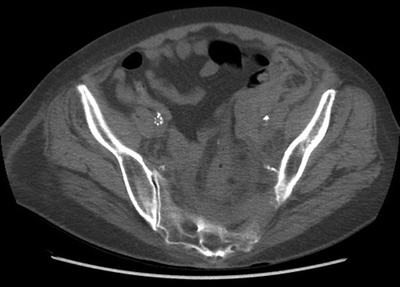
FIGURE 1 Pelvic CT.


