Restrictive Cardiomyopathy

20 Restrictive Cardiomyopathy


Cardiomyopathies are generally classified into three forms: dilated, hypertrophic, and restrictive. The restrictive form is the least common endomyocardial disease and is characterized by variable degrees of diastolic dysfunction out of proportion to systolic dysfunction. Clinically, restrictive cardiomyopathy is often and easily confused with constrictive pericarditis. Differentiating between them is a challenge but very important because of the implications for prognosis and treatment. Restrictive cardiomyopathy and constrictive pericarditis can both be present in the same patient, further complicating the diagnosis and therapeutic decision making. Because constrictive pericarditis is eminently more treatable than restrictive cardiomyopathy, the distinction is critical.


Restrictive cardiomyopathy was originally described in 1961 as constrictive cardiomyopathy. This was later changed to the more accurate term, restrictive cardiomyopathy, which describes a stiff myocardium usually resulting from an infiltrative process. Diastolic heart failure is now recognized to be a common process, often affecting the elderly and those with hypertension and increased systemic arterial stiffness. Although the etiology for diastolic dysfunction is restrictive cardiomyopathy in some patients, more commonly diastolic dysfunction and diastolic heart failure arise from other causes.


Etiology and Pathogenesis


A variety of disease states produce the clinical manifestation of a restrictive cardiomyopathic process (Box 20-1). Myocardial fibrosis, myocardial infiltration by specific proteins, endomyocardial scarring, and cardiac muscle hypertrophy all may present as diastolic dysfunction.


Noninfiltrative Causes


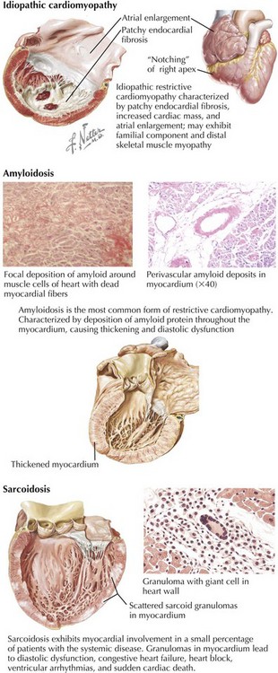
Idiopathic restrictive cardiomyopathy is associated with patchy endomyocardial fibrosis, increased cardiac mass, and enlarged atria (Fig. 20-1). It is more common in older adults but may be seen in children. In adults, 5-year survival is approximately 64%, but mortality may be higher in children. Occasionally, the cardiomyopathy is accompanied by skeletal muscle myopathy, and in some patients with restrictive cardiomyopathy, a clear familial component is present. Idiopathic restrictive cardiomyopathy is also found in families with no skeletal muscle involvement, however, and as an autosomal-dominant disorder in patients with Noonan’s syndrome. Conduction system disease such as atrioventricular (AV) block may also be present and in some cases precedes clinical myocardial dysfunction.


Infiltrative Causes


Clinically, the most common variety of restrictive cardiomyopathy is from amyloidosis, the deposition of unique, twisted, β-pleated sheets of fibrils formed by various proteins (Fig. 20-1, middle). Cardiac amyloidosis can be present in several different circumstances. Primary amyloidosis is caused by deposition of an amyloid protein composed of portions of an immunoglobulin light chain (designated AL for light chain–associated amyloidosis) produced by a monoclonal population of plasma cells. Primary amyloidosis can be the consequence of multiple myeloma but it is also found in patients without multiple myeloma. Secondary amyloidosis, sometimes called reactive systemic disease, is caused by the production of a nonimmunoglobulin protein and termed AA (for amyloid-associated). Familial amyloidosis is an inherited autosomal-dominant trait resulting from a variant prealbumin protein, transthyretin. More than 80 point mutations have been described, and familial amyloidosis may present as a cardiomyopathy, a progressive neuropathy, or a nephropathy. It is four times more common in blacks than in whites. In some cases, the heart is the only affected organ. Senile systemic amyloidosis is produced by an atrial natriuretic-like protein or transthyretin. Its frequency increases with age. Scattered amyloid deposits in the aorta or the atria are almost universally found in individuals older than 80 years, whereas only a small minority of the elderly has evidence of restrictive cardiomyopathy due to amyloidosis.


Regardless of the specific etiology, the overall size of the left ventricular (LV) chamber is normal or small, and at least early in the disease, systolic function is preserved even in individuals with very significant diastolic dysfunction. The greater the myocardial thickness, the more amyloid present and the worse the prognosis.


In amyloidosis secondary to immunocyte dyscrasias, cardiac involvement is common and the most frequent cause of death. In amyloidosis secondary to other diseases, cardiac involvement is much less common, often only manifesting as smaller perivascular deposits that do not cause diastolic dysfunction. About one in four individuals with familial amyloidosis has overt cardiac involvement, and even in these individuals, neurologic and renal dysfunction often dominate the clinical picture. Senile amyloidosis is rarely responsible for clinical cardiac dysfunction.


Symptomatically, patients with cardiac amyloid present with severe diastolic dysfunction and predominantly right-sided heart failure. Late in the course there may be progressive loss of LV systolic function and pulmonary congestion. Amyloid deposits in the atria are demonstrated by a markedly thickened interatrial septum and loss of atrial function. Most patients also experience arrhythmias and conduction system disease. Peripheral neuropathy is common, and orthostatic hypotension may be a major feature. Orthostasis is worsened by amyloid involvement in the adrenals and nephrotic syndrome due to renal involvement. Syncope is a common and ominous symptom.


Sarcoidosis is a granulomatous disease of unknown cause (Fig. 20-1, lower). Of the multiple organ systems commonly involved, including the heart, the most important is usually the lungs, where this involvement manifests as diffuse scarring, pulmonary hypertension, and cor pulmonale. Myocardial involvement causes a restrictive or a dilated cardiomyopathy in less than 5% of systemic sarcoidosis patients. More commonly, focal involvement may result in heart block, congestive failure, ventricular arrhythmias, or sudden cardiac death. The noncaseating granulomas have a propensity for involving the interventricular septum (hence the high incidence of heart block) and the LV free wall. The scattered nature of granulomas contributes to the failure of right ventricular (RV) biopsies to detect the disease in about half the patients. MRI is much more sensitive for detection of cardiac involvement in patients with known sarcoidosis.


Clinically, patients with sarcoidosis generally present with syncope from conduction system disease or cor pulmonale from both the pulmonary manifestations and cardiac involvement. Myocardial involvement may be gradually progressive, although it can be fulminant and lead rapidly to death.


Endomyocardial Causes


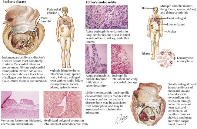
Endomyocardial fibrosis (sometimes called Becker’s disease) occurs most commonly in Africa, especially in Uganda and Nigeria (Fig. 20-2, left). In equatorial Africa, it is responsible for 10% to 20% of deaths from heart disease. Pericardial effusions are common and may be large. Fibrous endocardial lesions are frequently noted in the ventricular inflow tracts and often involve the AV valves, resulting in valvular regurgitation. The involved myocardium demonstrates a thick layer of collagen tissue overlying a layer of loosely arranged connective tissue. Fibrous and granulomatous tissue may extend into the myocardium. Either or both ventricles may be involved, and when the disease process is extensive, papillary muscles and chordae may be matted with a mass of thrombus and tissue, filling the cavity. Clinical manifestations depend on the extent of involvement of the right ventricle, the left ventricle, or both. Eosinophilic endocarditis (Löffler’s endocarditis) is probably an earlier manifestation of this same process (Fig. 20-2, lower). Both diseases are associated with eosinophilia. Epidemiologic evidence suggests that Löffler’s endocarditis is related to worm (helminth) infestation. It is thought that it is during the initial (necrotic) phase of hypereosinophilia that myocardial damage occurs. This is then followed after a year or more by a thrombotic phase and finally a fibrotic, restrictive phase. Clinically, the initial phase is characterized by fever, weight loss, rash, and congestive heart failure (CHF). Localized thickening of the posterolateral LV wall and limited mitral valve movement may be noted. In some instances, the LV apex is virtually obliterated by thrombus. Later, a restrictive pattern with AV regurgitation dominates the hemodynamics, and pericardial effusions, sometimes quite large, are seen.


Patients with Churg-Strauss syndrome (asthma, eosinophilia, neuropathy, pulmonary infiltrates, paranasal sinus abnormalities, and/or extravascular eosinophils) may also develop endomyocardial fibrosis. The intracytoplasmic granular content of activated eosinophils may be toxic to the myocardial and endothelial cells, resulting in the damage observed.


Prior radiation therapy is an important cause of restrictive cardiomyopathy. It is believed that radiation may cause long-lasting injury to the capillary endothelial cells, leading to cell death, capillary rupture, and microthrombi. Cardiac complications usually occur many years after the initial insult and can vary widely, with constrictive pericarditis a more common manifestation than restrictive cardiomyopathy. Pericarditis with effusion, coronary artery fibrosis (especially ostial) with myocardial infarction, valvular stenosis or regurgitation, conduction system disease, and myocardial fibrosis may result from excessive radiation exposure. The severity of cardiac involvement is proportional to the radiation dose (more common at doses greater than 45 Gy) and to the mass of myocardium exposed. Cardiac radiation exposure is most common following therapy for Hodgkin’s disease or breast cancer and, despite attempts to shield the heart from radiation, is still a concern. Additionally, although damage to the myocardium from chemotherapy (which many of these same patients receive) ultimately causes systolic dysfunction, diastolic dysfunction can be present. Separating the effects of radiation from the consequences of chemotherapy is not always possible.


The most common cardiotoxic chemotherapeutic agents are the anthracyclines. After anthracycline exposure, cardiac toxicity usually is delayed and results in a dilated cardiomyopathy. Early manifestations of primarily diastolic dysfunction may herald the cardiotoxicity. There is a nonlinear increase in cardiotoxicity as the cumulative dose increases, with a 7% incidence with doxorubicin doses over 550 mg/m2. Cytotoxicity from anthracylines seems to be due to the inhibition of an enzyme necessary for DNA repair and to generation of free radicals that damage cell membranes, in part by lipid peroxidation. The heart may not detoxify the free radicals because only a small amount of catalase, needed to convert hydrogen peroxide to water, is present. The anthracyclines also chelate iron and generate tissue-damaging hydroxyl radicals locally. Therefore, dexrazoxane, a drug that hydrolyzes to form a carboxylamine capable of removing the iron from the anthracycline-iron complex, is often used as a cardioprotectant in patients receiving anthracyclines. Other toxic drugs that have been implicated in the development of myocardial fibrosis include methysergide, ergotamine, mercurial agents, and busulfan.


Other Causes


Less common causes of restrictive cardiomyopathy include certain inherited diseases. The most prominent is Fabry’s disease, an X-linked recessive disorder caused by deficiency of the lysosomal enzyme α-galactosidase. The accumulation of lysosomal glycolipids in cardiac tissue results in a severe restrictive cardiomyopathy. Some patients with Fabry’s disease also have involvement of the cardiac valves, the skin, the kidneys, and the lungs.


Hypertrophic cardiomyopathy can present in a similar manner as a restrictive cardiomyopathy. Many mutations in sarcomeric proteins have been identified in genetic studies of hypertrophic cardiomyopathy (see Chapter 19), and there is variability in the degree of diastolic dysfunction depending on both the genotype as well as concomitant diseases (hypertension, diabetes). Generally, it is not difficult to distinguish hypertrophic cardiomyopathy from other causes of restrictive cardiomyopathy.


Other inherited diseases are rare and, hence, less commonly a cause of restrictive cardiomyopathy. In Gaucher’s disease (characterized by a deficiency of the enzyme β-glucosidase, with accumulation of cerebrosides in various organs), there may be both myocardial dysfunction and hemorrhagic pericardial effusion. In Hurler’s syndrome, a deposition of mucopolysaccharide in the myocardium can cause a restrictive process. The cardiac valves and the coronary arteries may also be involved. Hemochromatosis, arising from inherited (autosomal-recessive) or acquired etiologies, is characterized by iron deposition in many organs, including the heart. Myocardial damage may result from direct tissue damage by the free-iron moiety, not from the infiltration of iron. Many reports have described massive trabeculations in the left ventricle toward the apex with large sinus recesses between the trabeculae—a pattern that defines ventricular noncompaction. Noncompaction is a genetic disorder that may present with any or all of the features of a restrictive cardiomyopathy. Cardiac MRI is usually definitive.


Carcinoid heart disease primarily affects the right heart and is characterized by fibrous plaque that virtually coats the tricuspid and pulmonic valves and the RV endocardium. Valvular stenosis and regurgitation result, and RV dysfunction is common. The cardiac involvement in patients with carcinoid correlates with serotonin concentrations.


Differential Diagnosis


Most patients present with right heart failure out of proportion to left heart failure and have normal or near-normal cardiac size on examination and chest x-ray. Though not specific for restrictive cardiomyopathy, this constellation of symptoms, signs, and findings should always raise the possibility of restrictive cardiomyopathy. The differential diagnosis of restrictive cardiomyopathy includes several cardiac causes: constrictive pericarditis, chronic RV infarction, RV dysfunction from RV pressure or (less likely) RV volume overload, intrinsic RV myocardial disease, or tricuspid valve disease. Additionally, discerning restrictive cardiomyopathy from primary hepatic causes, including cirrhosis, can be challenging, since both can present with evidence of right heart failure, ascites, and marked hepatic dysfunction. Upon further evaluation, however, the results of the examination and echocardiography usually narrow the differential diagnosis to restrictive cardiomyopathy and constrictive pericarditis. These two entities affect hemodynamics in a subtly different manner, and distinguishing the two can be challenging but is extremely important given the prognosis and treatment options.


Normal Hemodynamics


Intracardiac pressures are a reflection of the contraction and relaxation of individual cardiac structures and the changes imparted to them by the pleural and pericardial pressures (Fig. 20-3). Changes in either pleural or pericardial pressure can be reflected in the intracardiac pressure. With inspiration, the intrapleural pressures drop and the abdominal cavity pressure increases. Blood flow through the right side of the heart increases, whereas blood return to the left side of the heart decreases slightly. The fall in the intrapleural pressures with inspiration also increases the transmural aortic root pressure, effectively increasing the impedance to LV ejection. The reverse occurs during expiration. Normally, inspiration lowers the right atrial and the systolic RV pressures slightly more than it lowers the left heart pressures. In severe lung disease, such as asthma, left heart filling is more profoundly affected, and these changes are exaggerated. The very negative inspiratory intrapleural pressures and very positive expiratory pressures result in marked swings in LV filling. A paradoxical pulse (fall in systemic pressure with inspiration) may thus result from lung disease alone.
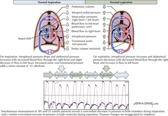


The normal atrial and ventricular waveforms are shown in upper Figure 20-4. With atrial contraction, the atria become smaller and the atrial pressures rise (a wave). With the onset of ventricular contraction, the AV valves bulge toward the atria, and a small c wave is typically detectable on hemodynamic tracings. Although many findings can be seen by careful inspection of the jugular veins on physical examination, the c

Only gold members can continue reading. Log In or Register to continue

Stay updated, free articles. Join our Telegram channel

Jun 12, 2016 | Posted by in CARDIOLOGY | Comments Off on Restrictive Cardiomyopathy

Full access? Get Clinical Tree

Get Clinical Tree app for offline access