Figure 43.1
Skin and greater pectoral muscle flaps are raised 5–6 cm lateral to the sternal border
Sternal Reconstruction
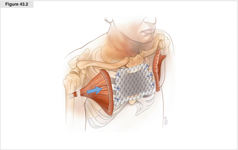
Several prosthetic materials are available to reconstruct sternal defects. Characteristics influencing the choice of a specific prosthetic include ease of use; flexibility; malleability to adapt to various sizes, shapes, and contours; durability; inertness to body fluids; resistance to infection; and incorporation by body tissues. Other important factors include the ability to provide rigid stability to the chest wall and to minimize paradoxic chest wall movement. In the end, the choice of prosthetic material is based on the surgeon’s preference, because all these materials work reasonably well. Since the mid-1980s, 2-mm thick polytetrafluoroethylene (PTFE; Gore-Tex, W. L. Gore and Associates, Inc., Flagstaff, AZ) has been our prosthesis of choice for reconstruction of all chest wall defects, including sternal reconstruction (Deschamps et al. 1999). PTFE is flexible, durable, and easy to suture, stretch, and mold into the wound. It is impervious to air and fluid and provides a barrier that prevents air and fluid from moving between the pleural and subcutaneous spaces. We secure the PTFE patch with heavy interrupted nonabsorbable monofilament sutures (Prolene, Ethicon, Somerville, NJ). The sutures are placed either through or around the ribs (Fig. 43.2). We prefer to use a handheld pneumatic drill to create 2-mm holes in the ribs to ensure secure suture placement. Once all the sutures have been placed, care should be taken to stretch the patch taught as the sutures are tied down.
An alternative technique is to use polypropylene mesh (Marlex, Bard Cardiosurgery, Billerica, MA). Marlex is a single-stitch knit mesh that is malleable in one direction and rigid in the other. It is a semipermeable membrane with a pore size of 75 μ. The mesh enables macrophages and fibroblasts to pass through and provides a matrix for tissue ingrowth, allowing polypropylene to be incorporated by the surrounding tissue. Proponents of polypropylene mesh claim that it is less likely to engender an overlying seroma, which may lead to fewer wound infection problems. When we retrospectively reviewed the early and long-term results of 197 patients who underwent chest wall resection and reconstruction using either polypropylene mesh or PTFE, we found no difference in the rates of seromas, wound infections, or other postoperative complications between the two prosthetic materials (Deschamps et al. 1999).
The use of methylmethacrylate in conjunction with polypropylene mesh was described initially by McCormack and colleagues at Memorial Sloan-Kettering Cancer Center in 1981 (McCormack et al. 1981). Since then, the technique of using a sandwich of two layers of Marlex mesh with methylmethacrylate between them has been widely adopted in sternal reconstruction because of its ability to provide a rigid protective plate covering the heart and other mediastinal viscera. Once the sternum has been resected, the specimen is placed in the middle of a sheet of Marlex mesh and an outline of the sternum is traced onto the mesh as a template for fashioning the methylmethacrylate prosthetic. The methylmethacrylate powder is mixed with the solvent, and the resulting paste is placed on top of the mesh following the outlines of the sternal defect (Fig. 43.3). A second layer of Marlex is placed on top of the methylmethacrylate, and the composite sandwich is allowed to harden. Before the polymerization of the methylmethacrylate is complete, it is easy to mold and contour it to the exact size and shape of the defect. Care must be exercised during the hardening phase because of the exothermic nature of the polymerization reaction. Temperatures may reach 140 °F, causing surrounding tissue necrosis if the composite sandwich is not allowed to cool before being placed in the wound. A useful technique is to place the Marlex–methylmethacrylate sandwich onto a thin sheet of malleable lead. This allows the composite prosthetic to be molded to the precise convexity desired while it is still in its semisolid state and also provides a scaffold that can tolerate the exothermic polymerization phase. Excess mesh is then trimmed from the composite, leaving a 2-cm rim of mesh as a “sewing ring.” Heavy nonabsorbable monofilament suture is used to secure the prosthetic to the skeletal defect (Fig. 43.3).


