Renal Vein Entrapment: The Nutcracker Syndrome


Renal Vein Entrapment


The Nutcracker Syndrome


Brandon T. Garland and Mark H. Meissner


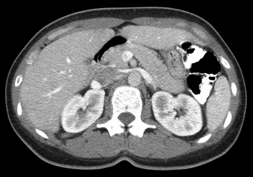
The nutcracker syndrome is characterized by entrapment and compression of the left renal vein between the abdominal aorta and the superior mesenteric artery (SMA) (Figure 1). The underlying anatomic anomaly, first described by Gant in 1937, can lead to left renal venous hypertension and variable degrees of hematuria and left flank pain. In women, it may be associated with pelvic congestion symptoms, including pelvic pain, dyspareunia, dysuria, and gluteal, perineal, and labial varices; manifestations in men can include a varicocele. Not all patients with compression identified by noninvasive imaging have symptoms, and among those with trivial symptoms, many can be managed conservatively. However, for those with disabling symptoms, surgical or endovascular treatment can provide substantial relief.


Anatomic Features


The left renal vein most commonly follows a course between the SMA anteriorly and the aorta posteriorly. Symptomatic compression of the renal vein between the SMA and aorta is termed anterior nutcracker syndrome and has been attributed to a variety of causes including an acute or lateral origin of the SMA, fibrosis surrounding the SMA origin, a high trajectory of the left renal vein, and posterior ptosis of the left kidney. Most attention has focused on the origin of the SMA, which normally arises from the aorta at a near-90-degree angle and courses ventrally for 4 to 5 mm to form a rectangular tunnel through which the left renal vein passes. Both the third portion of the duodenum and retroperitoneal fat are postulated to be of importance in maintaining a wide aortomesenteric angle.


The SMA has been found to arise at a much more acute angle in many patients with the nutcracker syndrome. However, there is wide variability in both the distance between the aorta and SMA and the aortomesenteric angle, and it remains unclear if either of these measurements predict a hemodynamically significant renal vein compression. The left renal vein can also course posterior to the abdominal aorta (retroaortic or circumaortic renal vein), allowing compression between the aorta and vertebral column, referred to as the posterior nutcracker phenomenon. Finally, the left renal vein can be compressed by pancreatic and other retroperitoneal neoplasms, an aberrant testicular artery, or paraaortic lymphadenopathy, all of which encroach into the aortomesenteric space.


Regardless of etiology, central obstruction of the left renal vein can lead to renal venous hypertension with the development of renal pelvic varices and collateral drainage through tributaries. Important tributaries that can function to drain blood exiting the kidney include the left adrenal, gonadal, ascending lumbar, hemiazygous, periureteric, and capsular veins. The adequacy and specific anatomy of this collateral drainage likely determines the clinical manifestations of the nutcracker syndrome.


The gonadal veins are among the most important collateral pathways, and differences in anatomy might account for the gender specificity of many symptoms. In women, the ovarian vein communicates with tributaries of the internal iliac veins. Reflux in these systems can lead to pelvic congestion symptoms including pelvic pain, dysuria, and dyspareunia. It can also lead to atypical varices (labial, perineal, or gluteal) as well as to lower extremity varices if pelvic reflux reenters the saphenous vein through external pudendal tributaries. In men there is no collateral flow through the pelvis, and gonadal vein reflux can lead to a left-sided varicocele.


Clinical Features


Asymptomatic compression of the left renal vein, often termed the nutcracker phenomenon, must be distinguished from hemodynamically significant compression associated with clinical symptoms, the nutcracker syndrome. In comparison to the hilar segment, between 10.9% and 72% of patients have evidence of at least 50% compression of the aortomesenteric segment of the renal vein on noninvasive imaging. Among patients with signs of compression on computed tomography (CT) imaging, Poyraz found that only 8.8% had hematuria or proteinuria, and only 5.5% had a varicocele or pelvic congestion symptoms. Similarly, renal vein varices were identified in only 10% of consecutive left renal venograms, and symptoms were present in only one quarter of these patients. Asymptomatic nutcracker phenomenon is therefore much more common than symptomatic nutcracker syndrome.


The development of symptoms appears slightly more common in women. Some have suggested a bimodal age distribution with one peak in adolescents and young adults and a second in women in their third to fourth decades. Spontaneous resolution of symptoms may be more common in younger patients. The syndrome has also been reported to be more common in asthenic patients, presumably because of a paucity of retroperitoneal fat associated with either narrowing of the aortomesenteric angle or posterior ptosis of the kidney. A moderate correlation has been observed between body mass index (BMI) and the aortomesenteric angle determined by ultrasound. There is no known hereditary predisposition to the disorder.


The nutcracker syndrome is classically associated with left flank pain and hematuria, which may be absent or intermittent and either gross or microscopic. Such symptoms are often exacerbated in the upright position and relieved with recumbency. Hematuria, and associated orthostatic proteinuria, is postulated to result from rupture of thin-walled perirenal varices into the collecting system. However, the nutcracker syndrome may also be associated with an array of pelvic and lower extremity symptoms in the absence of hematuria. Symptoms of pelvic congestion syndrome with or without perineal and lower extremity varies in women, and a left-sided varicocele in men may be noted. Approximately 90% of varicoceles are on the left side, and many patients demonstrate distinct ultrasound findings consistent with renal vein compression. Given the spectrum of associated symptoms, it is not surprising that many of these patients undergo extensive gynecologic or urologic investigation before they are seen by a vascular specialist.

Stay updated, free articles. Join our Telegram channel

Aug 25, 2016 | Posted by in CARDIOLOGY | Comments Off on Renal Vein Entrapment: The Nutcracker Syndrome

Full access? Get Clinical Tree

Get Clinical Tree app for offline access