Renal Ectopia and Renal Fusion in Patients Requiring Abdominal Aortic Operations


Renal Ectopia and Renal Fusion in Patients Requiring Abdominal Aortic Operations


Patrick J. O’Hara


Renal ectopia is a developmental anomaly in which the position of one or both kidneys is abnormal. It can occur alone or in combination with fusion or malrotation of both renal masses. Congenital anomalies of the upper urinary tract are among the most common organ system anomalies encountered by the vascular surgeon, and they may also commonly be associated with coexisting anomalies of renal vasculature and the collecting system. The reported prevalence of renal ectopia ranges from 1 in 500 to 1 in 1200 (average, 1 in 900) in autopsy series. Horseshoe kidney is the most common renal fusion anomaly, occurring in 0.25% of the population or about in 1 in 400 persons. Because the presence of these renal anomalies can complicate abdominal aortic operations, an appreciation of the pertinent embryology is helpful to visualize the anatomic changes in such patients.


Embryology


Three renal precursors, called the pronephros, the mesonephros, and the metanephros, develop from the mesoderm during early gestation. The first two of these early structures regress, but around the 28th day of gestation, the metanephros differentiates into the fetal kidney, which forms in the sacral region. In this primitive structure, a pair of ureteric buds develops and interacts with the metanephric mesenchyme to form a pair of fetal kidneys. During this process, the collecting system develops from each ureteric bud, and the nephrons develop from the metanephric mesenchyme through reciprocal interaction. Between the 6th and 9th weeks of gestation, the two kidneys rotate and ascend from the pelvis to their usual anatomic position in the right and left side of the lumbar region, just below the adrenal glands. As the kidneys ascend, they receive their blood supply through a series of paired arteries arising from the primitive aorta, which sequentially disappear as the kidneys migrate. The result is usually a single pair of renal arteries, one for each kidney. Each renal segment is usually supplied by a single end-arterial branch originating from the main renal artery, but between 15% and 30% of kidneys have more than one renal artery.


If the embryonic kidneys fuse during development, normal rotation and ascent are interrupted, leading to a variety of anomalies ranging from positional disorders alone to a combination of fusion, ectopia, and malrotation problems. Although isolated renal artery anomalies are usually easily handled by the experienced vascular surgeon, renal parenchymal developmental abnormalities are less common and are often associated with anomalies of the collecting system as well as the vasculature. The combination can pose a technical challenge to the vascular surgeon at the time of aortic reconstruction, especially during repair of abdominal aortic aneurysm (AAA), and may be particularly difficult to manage if it is encountered unexpectedly, because adequate exposure may be difficult through the traditional anterior transperitoneal approach to the infrarenal aorta. The risk of injury to the urinary collecting system may be increased because of the anterior location of the renal mass, and ligation of anomalous renal arteries can lead to renal infarction. For these reasons, preoperative imaging, when feasible, is valuable at the outset to allow the preoperative detection of associated renal anomalies and to plan the most advantageous choice of incision and patient positioning.


Anatomic Subgroups of Renal Ectopia


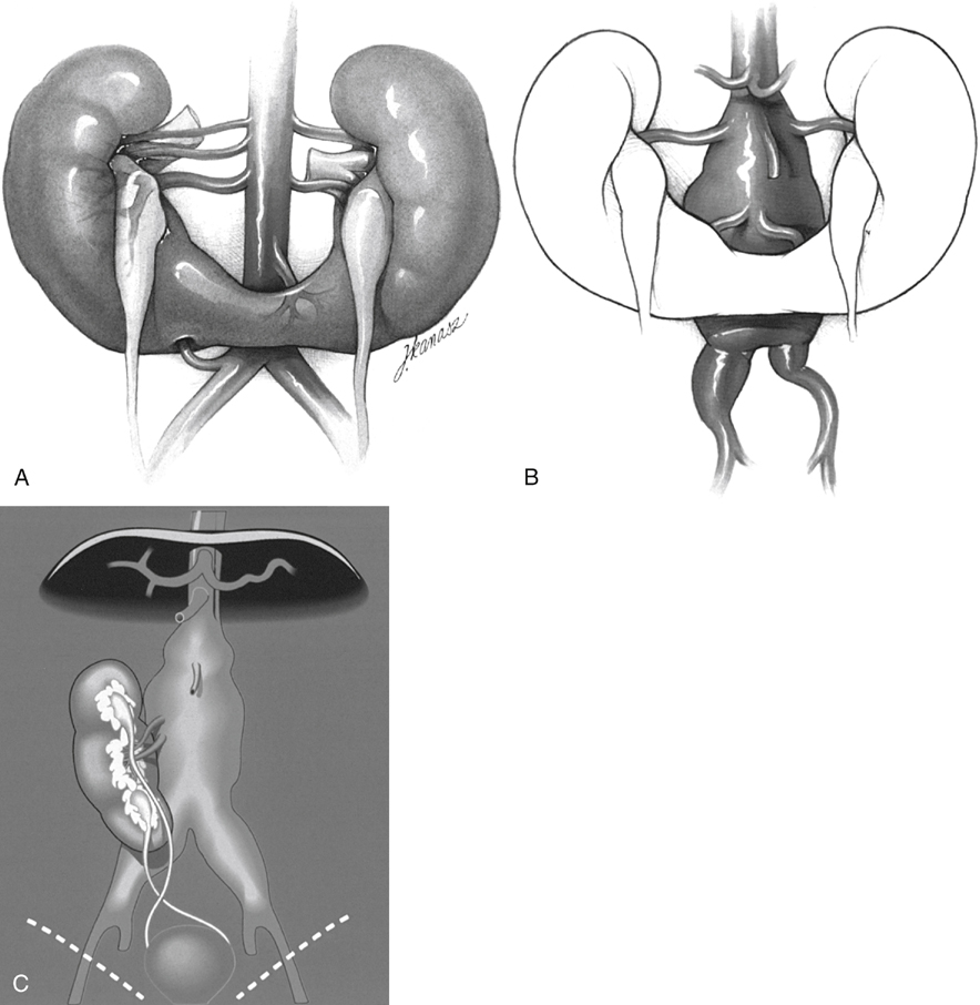
Developmental anomalies of the kidneys and upper urinary tracts may be categorized according to those leading to problems of number, ascent, rotation, or form and fusion of the renal parenchymal mass (Box 1). All may be associated with abnormalities in the renal vasculature as well as in the collecting system, but the problems with renal form and fusion are typically the most troublesome for the vascular surgeon during AAA repair. Anomalies of renal ascent or rotation alone rarely seriously interfere with traditional approaches to aortic reconstruction. In contrast, anomalies of fusion are usually associated with varying degrees of ectopia and malrotation, which can require modification of standard surgical techniques.


Horseshoe Kidney


Horseshoe kidney, thought to occur in 0.25% of the population, is probably the most common of all renal fusion abnormalities and is twice as common in men as it is in women. Horseshoe kidneys result when the metanephric masses fuse anteriorly in the midline. Their normal bilateral ascent is presumed to be blocked by the inferior mesenteric artery, and the associated impaired medial orientation causes the renal hila and collecting systems to be rotated ventrally, in turn inducing the ureters to lie anterior to the renal isthmus (Figure 1A).


The horseshoe kidney usually consists of two separate renal masses lying vertically on either side of the midline and connected at their respective lower poles by an isthmus that crosses anterior to the aorta near the origin of the inferior mesenteric artery. This can pose a particular challenge during AAA repair (see Figure 1B). Although occasionally fibrous and avascular, the isthmus may consist of substantial renal parenchyma and its blood supply may be asymmetric, making simple division of the isthmus in the midline hazardous. Furthermore, the collecting systems may also be asymmetric and can extend anteriorly and across the midline in the region of the isthmus. Arterial and venous anomalies are common.


Associated arterial anomalies occur in 60% to 80% of horseshoe kidneys. In the author’s series, renal artery anomalies were encountered in 14 patients (74%), whereas the remaining five patients (26%) had only two main renal arteries in the normal location. Division of the renal isthmus was avoided in all patients of the author’s series to avoid the risk of urinary fistula and graft sepsis, because 13% of patients with horseshoe kidney are reported to have chronic urinary tract infection.


Crossed Renal Ectopia


If the fused metanephric masses ascend together on one side, in contrast to horseshoe kidney development, then crossed renal ectopia can result. In this anomaly, the ectopic kidney is located on the side contralateral to its ureterovesical junction and, in most instances, is fused to the inferior portion of the ipsilateral kidney (see Figure 1C). Fusion and crossover of the left kidney to the right side is the most common form of this variation and results in a single fused renal unit, with the collecting system lying anterior to the aorta and at least one of the ureters crossing the midline anterior to the aorta, usually above the second sacral vertebra. The arterial and venous anatomy often consists of multiple vessels in unpredictable locations. Crossed renal ectopia has been further classified depending on the shape assumed by the fused renal masses (see Box 1), but all forms are managed the same way during aortic reconstruction.

Only gold members can continue reading. Log In or Register to continue

Stay updated, free articles. Join our Telegram channel

Aug 25, 2016 | Posted by in CARDIOLOGY | Comments Off on Renal Ectopia and Renal Fusion in Patients Requiring Abdominal Aortic Operations

Full access? Get Clinical Tree

Get Clinical Tree app for offline access