Renal Artery Dissection


Renal Artery Dissection


Linda M. Reilly


Renal artery dissection is most commonly associated with dissection of the aorta. Less than one fourth of all dissections involving the renal vessels are isolated to the renal artery alone. Nonetheless, the renal artery is the most common site of isolated dissection of an aortic branch vessel.


The causes of isolated renal artery dissection fall into two distinct categories: those that are primary and associated with underlying renal artery diseases such as fibromuscular dysplasia or atherosclerosis, and those that are secondary and a consequence of blunt trauma or interventional procedures. The clinical sequelae of isolated renal artery dissection depend upon the degree of reduction of renal blood flow resulting from the compromised true lumen, varying from clinically silent, to hypertension caused by renal ischemia, to frank renal infarction. The approach to treatment depends upon the clinical setting and the extent of the anatomic defect in the renal artery.


Endovascular Procedure-Induced Dissection


Dissection is a potential complication of endoluminal instrumentation of the renal artery. The risk of dissection increases as the complexity of instrumentation progresses from simple selective renal artery catheterization for diagnostic imaging, to balloon angioplasty, and stent or covered-stent insertion. It is difficult to establish an accurate incidence of this complication because endpoint definitions vary and reporting standards have not been established. Therefore it is likely that many small, limited, clinically silent dissections resulting from endovascular procedures are not reported. Nevertheless, studies from about a decade ago reported rates of dissection less than 1% with selective renal angiography, 2% to 5% with renal artery angioplasty, and 5% to 7% with renal artery stenting.


The risk of endovascular procedure–induced dissection is related to the anatomy of the renal artery (diameter, tortuosity) and the nature of the underlying disease. In comparison to other aortic branches, the renal arteries are smaller in caliber, shorter, more mobile, and more tortuous in both the craniocaudal and anteroposterior planes. Each of these anatomic features increases the risk of dissection during instrumentation. Other anatomic features that influence the risk of dissection include aortic tortuosity (impairs tracking of catheters and devices) and prior pararenal aortic or renal artery surgical procedures (scar tissue fixation prevents the renal arteries from deforming to accommodate the catheters and delivery systems). Dissection is also more likely to occur during instrumentation of arteries with complex atherosclerotic lesions, but it is relatively less likely to occur during instrumentation of arteries involved by fibromuscular dysplasia (FMD). In reviewing 22 reported cases of endovascular procedure–induced renal artery dissections, Smith and coworkers noted associated atherosclerosis in 44% and FMD in only 18%.


Endovascular procedure-related renal artery dissection is usually unilateral because it is confined to the instrumented artery, involves renal artery branches in only about one quarter of patients, and has an equal gender distribution. In contrast to post-trauma and spontaneous dissections, endovascular procedure-related dissection is generally diagnosed immediately during the procedure that caused the dissection. About a third of the recognized instrumentation-related renal artery dissections are asymptomatic, and pain and hypertension occur in about 50% of the patients, which is a much lower rate than is seen in primary dissections. Hematuria occurs in about 20% of patients.


In the half of reported cases of dissection following renal artery intervention, the patients underwent renal artery reconstruction, usually involving a renal artery bypass. This approach is most successful when the dissection is confined to the main renal artery, when renal infarction has not yet occurred, and when revascularization can be accomplished quickly. Studies report patients treated with endoluminal techniques, which have the advantage of immediacy. Successful endoluminal treatment usually requires placing a stent or covered stent to obliterate the false lumen and reexpand the true lumen. In some cases, adjunctive thrombolysis is needed, particularly if partial or distal thrombosis or embolization has occurred. The key to successful endoluminal treatment is the ability to maintain access to the true lumen distal to the dissection and lack of involvement of segmental branches. Occasionally, particularly when dissection extends distally beyond what could feasibly be treated either with stents or operative reconstruction, endoluminal distal fenestration of the septum provides the only option for improving distal perfusion.


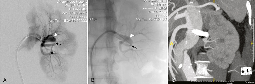
In early series, about one third of patients were observed, either because they were clinically stable (symptoms resolved, hypertension resolved or was medically controlled, and renal function remains normal or only minimally impaired) or because there were no evident treatment options. In this patient group, interval imaging often shows remodeling of the dissected artery, as well as some improvement in renal parenchymal perfusion (Figure 1), although complete normalization of arterial anatomy and renal perfusion is uncommon. Occasionally, improvement is unexpectedly dramatic. It is important to keep this in mind when determining how aggressive to be in attempting endoluminal treatment, which has the risk of worsening the dissection, or surgery, which has the risk of resulting in a nephrectomy. Although early series reported at least a 15% incidence of nephrectomy, it is important to avoid early nephrectomy whenever possible, because significant spontaneous improvement of the abnormal arterial anatomy and impaired renal perfusion can occur. Nephrectomy should be reserved for patients who develop refractory hypertension or other symptoms that suggest clinical instability.


Blunt Trauma–Related Dissection


Injury to the main renal artery by blunt abdominal trauma is a rare occurrence (estimated incidence of <1%) but is now recognized more often as a result of the common use of computed tomography (CT) to evaluate trauma victims. Direct impact is one mechanism of injury, but the mobility of the renal pedicle adjacent to the fixed aorta allows acceleration–deceleration injury and is probably more common. Because of the force needed to injure the renal artery, 60% to 80% of these patients have multiple associated injuries, contributing significantly to the reported 20% to 45% mortality rate.


Following blunt trauma, the most common finding on imaging studies is renal artery occlusion, but the underlying status of the renal artery can be difficult to determine from imaging alone. The occlusion can result from avulsion or transection, laceration, or dissection. Direct examination of the artery at surgery or selective imaging during endovascular treatment show that the underlying morphology is usually a dissection.


The management of dissection resulting from blunt abdominal trauma remains controversial. Treatment options include no intervention with interval imaging, endoluminal treatment, surgical revascularization, or nephrectomy. Attempts at surgical revascularization have produced disappointing results. Attempted renal artery revascularization was clinically appropriate in only about 25% of these patients; of those, only 25% regained any renal function and only about 10% of those had normal renal function. Delay in diagnosis and delay in treatment related to prioritizing the treatment of other injuries certainly contributes to these results. These outcomes, combined with the overall mortality in this patient group, have resulted in general agreement that unilateral renal artery dissection or occlusion after blunt abdominal trauma should not be treated surgically. Unfortunately, unlike endovascular procedure–related renal artery dissection, spontaneous recovery of kidney function in these patients is unusual, particularly if imaging shows no flow. Among patients treated with observation, more than half ultimately require nephrectomy.


It is not surprising that these results would prompt a search for alternative approaches. There have been several recent reports of endovascular renal revascularization following blunt abdominal renal artery injury. This approach has several potential benefits. It can be performed without adding further physiologic stress to a patient with multiple other injuries. It may be possible to achieve renal reperfusion with less delay, particularly if endovascular renal revascularization is performed in concert with interventional treatment of other associated injuries.


However this approach also has risks. Heparin anticoagulation cannot be used, increasing the risk of thrombus formation at arterial access sites, as well as on the wires, sheaths, catheters, and device-delivery systems. Manipulation of an injured artery can produce overt bleeding, converting a stable patient into an unstable one. The interventionalist must be prepared to quickly control any bleeding, whether this requires covered stent placement or occlusion of the artery with sacrifice of the kidney.


No large series of endoluminal treatment of blunt renal artery injury exist and therefore its role is uncertain. Anecdotal reports have established feasibility, but small series show a similarly disappointing low rate of renal function salvage of only 50%, with a significant rate of early occlusion or reocclusion of the stented artery.


Overall, it is the extent and duration of warm renal ischemia that determines the likelihood of success in salvaging kidney function following blunt renal artery trauma. Although evidence suggests that renal tissue can remain viable for up to 24 hours, most conclude that revascularization has little chance of success if it occurs after 4 hours of interrupted flow. Unfortunately, the delay following the injury (transport, diagnosis, stabilization, treatment) usually exceeds the tolerance of the kidney for warm ischemia. Beyond this window most surgeons will not undertake treatment for unilateral dissections related to blunt trauma. However if the injury is bilateral, most will undertake treatment up to 20 to 24 hours after injury. If there is still some flow through the injured renal artery, endoluminal treatment probably offers the best risk-to-benefit balance if any attempt to treat is undertaken. Future investigations should differentiate between patients presenting with and without residual renal perfusion and focus on determining the optimal treatment in that setting.

Stay updated, free articles. Join our Telegram channel

Aug 25, 2016 | Posted by in CARDIOLOGY | Comments Off on Renal Artery Dissection

Full access? Get Clinical Tree

Get Clinical Tree app for offline access