Renal Arteriovenous Malformations and Arteriovenous Fistulas


Renal Arteriovenous Malformations And Arteriovenous Fistulas


Kyung J. Cho and James J. Shields


Renal arteriovenous malformations (AVMs) and arteriovenous fistulas (AVFs) have the common characteristic of one or more abnormal channels between an intrarenal artery and vein, allowing the blood to bypass the glomerular capillaries. A communication between the renal artery and the renal vein can be demonstrated by color Doppler sonography, computed tomography (CT), or magnetic resonance angiography (MRA), and the diagnosis is confirmed by renal arteriography. The clinical significance, natural history and optimal therapy of the asymptomatic arteriovenous communications remain inadequately defined.


Classification


According to their pathogenesis, renal lesions with an abnormal communication between the renal artery and the renal vein can be classified as congenital AVMs (developmental) and acquired AVFs (traumatic, spontaneous, or neoplastic).


AVFs account for 70% to 80% of abnormal renal arteriovenous communications. Traumatic AVFs may be extrarenal or intrarenal. Extrarenal AVFs involve the main renal artery and renal vein and are often caused by nephrectomy or by penetrating injury. Intrarenal AVFs can involve the segmental, interlobar, and arcuate renal arteries and are most commonly caused by renal biopsy or by trauma, either blunt or penetrating. Spontaneous fistulas may be associated with renal artery disease, such as renal artery aneurysm and arterial fibrodysplasia.


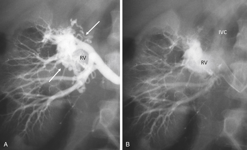
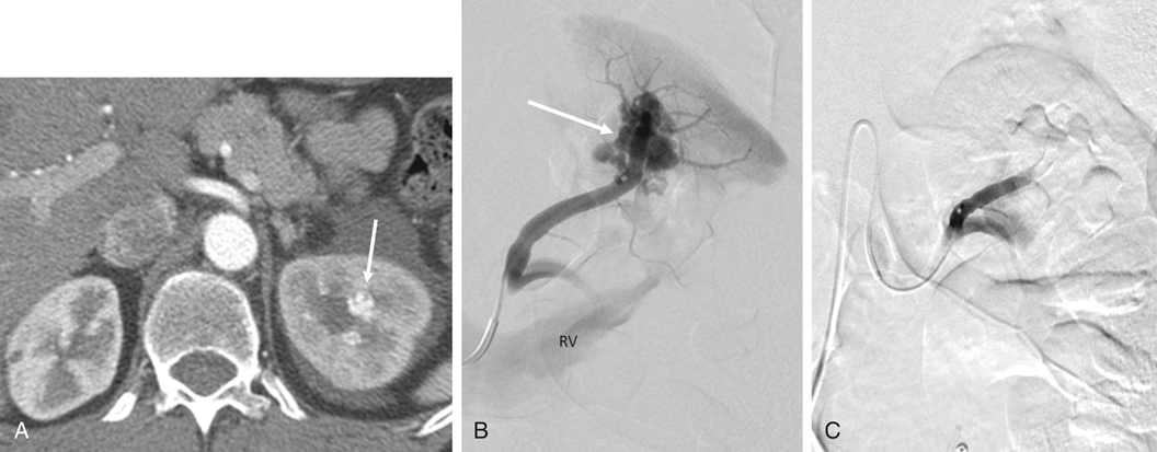
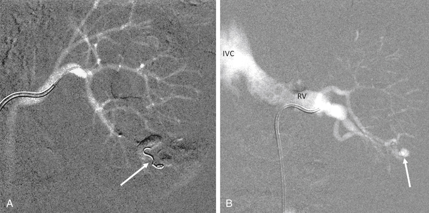
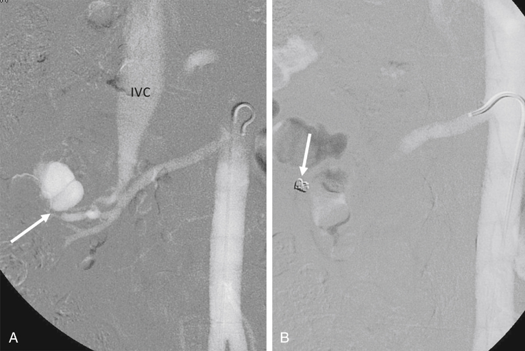
AVMs are congenital, are characterized by an arteriovenous communication at the arteriole and venule level, and are usually supplied by multiple feeding arteries (Figures 1 and 2). These malformations are either cirsoid, with multiple arteriovenous communications, or cavernous, with well-defined arterial and venous channels. In contrast, the acquired AVF usually has a single feeding artery supplying a direct communication between an artery and a vein (Figure 3).


Clinical Features


Clinical manifestations of renal AVMs and acquired AVFs depend on their size, location, and cause. Although nontraumatic and nonneoplastic AV communications are usually silent clinically, they have been alleged to cause hematuria, hypertension, spontaneous rupture, and heart failure. Symptomatic congenital AVMs usually manifest with hematuria varying in severity and uncommonly present with hypertension. Rarely, massive hematuria occurs with hypotension requiring blood transfusion. Hematuria is the most common presentation in traumatic fistulas. A large AVF can increase the size of the renal arteries and lead to high-output heart failure and renal insufficiency. Most patients with spontaneous AVFs associated with renal artery disease are hypertensive and rarely have hematuria. Decreased renal perfusion distal to the fistula is speculated to be a cause of renovascular hypertension, but a definite cause-and-effect relationship has not been well established.


Angiographic Diagnosis


Color Doppler sonography, CT, and MRA are useful in identifying abnormal renal AV communications. However, catheter-based angiography is the most accurate method for the diagnosis of AVMs and AVFs, and can provide a practical guide to effective treatment planning.


Arteriographic study should begin with an anteroposterior aortogram. The frame rates for the aortogram should cover the arterial phase at 3 to 4 frames per second. A flush catheter (5-Fr pigtail or 5-Fr Omni flush) should be placed so that the side holes are at the level of the renal arteries, and contrast medium should be injected at 15 to 20 mL/sec for 2 seconds. Carbon dioxide gas (CO2) can be used as an alternative contrast agent in the patient with contrast medium allergy, with kidney failure, or at high risk for contrast-induced nephropathy. Because of the low viscosity, CO2 is more sensitive in filling the AV fistula (see Figure 3).


Selective renal angiography should be performed to obtain detailed vascular anatomy and to find a cause for the patient’s symptoms before treatment. The flush catheter is exchanged over a guidewire for a curved-tip catheter such as a 5-Fr Cobra catheter or a 5-Fr shepherd’s hook catheter. Selective renal angiography is then performed in anteroposterior and oblique projections using magnification technique. The injection rate of contrast medium should be tailored according to the renal artery blood flow, usually 6 to 8 mL/sec. Because of the fast blood flow through the fistula, the acquisition frame rates should be at least 4 frames per second. If CO2 is used as the initial contrast agent, a renal arteriogram with iodinated contrast medium should be performed for detailed vascular anatomy before undertaking intervention such as selective arterial embolization.


For the evaluation of a renal transplant, pelvic arteriography should be performed before a selective renal arteriogram. Because of its buoyancy, CO2 is effective in identifying the renal artery and an AV communication in a transplanted kidney situated in a nondependent location in relation to the injection site in the iliac artery. Repeat arteriography with iodinated contrast medium should be obtained for a vascular roadmap before selective arterial embolization.


The angiographic abnormalities of renal AVMs and AVFs depend on the etiology, size, location, and extent of the lesions. Small, localized congenital AVMs have dilated, tortuous, and coiled vascular channels grouped in a cluster, which are supplied by multiple dilated branches from the segmental or interlobar arteries. The lesion is usually located in the hilar or pericalyceal region, ranging in size from 3 to 4 cm. They can occur in the upper polar, interpolar, or lower polar regions. Arteriovenous shunting, although often minimal, is always present and is demonstrated by early and dense filling of the vein. Like AVMs that occur elsewhere, neither capillary nor parenchymal contrast accumulation is present during the nephrographic phase (see Figure 1). Small polypoid defects of the calyces may be present.


Traumatic AVFs are usually solitary, with an easily identified feeding artery and draining vein (see Figure 3). These may be located in the renal medulla or parenchyma and can involve the segmental, interlobar, or arcuate arteries. Pseudoaneurysms occasionally coexist with traumatic fistulas (Figures 3 and 4). Spontaneous AVFs can occur in patients with renal artery aneurysms and renal artery fibrodysplasia. Renal angiography demonstrates any large aneurysm associated with an AVF. In such cases, the feeding artery and the draining vein are usually dilated. Arteriovenous shunting associated with arterial fibrodysplasia has been shown to be minimal and small in size.


Stay updated, free articles. Join our Telegram channel

Aug 25, 2016 | Posted by in CARDIOLOGY | Comments Off on Renal Arteriovenous Malformations and Arteriovenous Fistulas

Full access? Get Clinical Tree

Get Clinical Tree app for offline access