Recognition and Treatment of Abdominal Compartment Syndrome Following Ruptured Abdominal Aortic Aneurysm Repair


Recognition and Treatment of Abdominal Compartment Syndrome Following Ruptured Abdominal Aortic Aneurysm Repair


Dieter Mayer and Mario Lachat


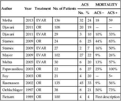
Abdominal compartment syndrome (ACS) and intraabdominal hypertension (IAH) caused by elevated intraabdominal pressure (IAP) are commonly encountered in patients with ruptured abdominal aortic aneurysms (AAAs) and carry a high morbidity and mortality risk (Table 1). However, IAH/ACS are still overlooked by many physicians and therefore timely diagnosis is not made and treatment is often inadequate.


Detection


The World Society of the Abdominal Compartment Syndrome (WSACS) has first brought order into the chaos of differing definitions of IAH and ACS by publishing part one of the WSACS Consensus Definitions and Recommendations in 2006. In this document, state-of-the-art definitions for IAH and ACS are proposed based upon current medical evidence as well as expert opinion. In 2013, these consensus definitions and recommendationshave been revised and resulted in updated consensus definitions and clinical practice guidelines (Box 1).


BOX 1   World Society of the Abdominal Compartment Syndrome Consensus Definitions


Definition 1: IAP is the pressure concealed within the abdominal cavity.


Definition 2: APP = MAP – IAP


Definition 3: Filtration gradient = GFP – PTP = MAP – 2 × IAP


Definition 4: IAP should be expressed in millimeters of mercury and measured at end expiration in the complete supine position after ensuring that abdominal muscle contractions are absent and with the transducer zeroed at the level of the midaxillary line.


Definition 5: The reference standard for intermittent IAP measurement is via the bladder, with a maximum instillation volume of 25 mL of sterile isotonic saline.


Definition 6: Normal IAP is approximately 5–7 mm Hg in critically ill adults.


Definition 7: IAH is defined by sustained or repeated pathologic elevation of IAP ≥12 mm Hg.


Definition 8: IAH is graded as follows:


Definition 9: ACS is defined as a sustained IAP >20 mm Hg (with or without an APP <60 mm Hg) that is associated with new organ dysfunction or failure


Definition 10: Primary ACS is a condition associated with injury or disease in the abdominopelvic region that often requires early surgical or interventional radiologic intervention.


Definition 11: Secondary ACS refers to conditions that do not originate from the abdominopelvic region.


Definition 12: Recurrent ACS refers to the condition in which ACS redevelops following previous surgical or medical treatment of primary or secondary ACS.


ACS, Abdominal compartment syndrome; APP, abdominal perfusion pressure; GFP, glomerular filtration pressure; IAH, intraabdominal hypertension; IAP, intraabdominal pressure; MAP, mean arterial pressure; PTP, proximal tubular pressure.


Routine surveillance for IAH and ACS should be implemented in patients treated for ruptured AAA in every intensive care unit, and a steady education program should be provided for all staff involved in managing those patients. Clinical examination alone has been shown to be inaccurate in estimating IAP. Hence, a variety of methods have been published regarding how to best measure or monitor abdominal pressure. Extensive research has shown that IAP can reliably be measured via the transvesical route by measuring either continuous or intermittent bladder pressure. One quick and simple way to assess IAP is to use an existing three-way Foley catheter (FoleyManometer, Holtech Medical, Charlottenlund, Denmark) connected to the patient’s urine catheter, using the patient’s own urine as a pressure medium (zero established at the iliac crest in the midaxillary line level). A higher incidence of urinary tract infections accompanying these instrumentations has not been proved.


The WSACS website (www.wsacs.org) provides several links to educational movies showing various techniques of measuring bladder pressure with different industrially assembled IAP measurement kits (see Education, IAP techniques [requires password]). It is generally regarded as less important which method is used for IAP measurement so long as IAP is routinely measured. IAP values should always be expressed as mm Hg (1 mm Hg = 1.36 cm H2O).


Unresolved issues remain, such as how much fluid should be instilled into the bladder to measure IAP, in which position IAP should be measured, and what the confounders would be for adequate IAP measurement. To date, a maximal instillation volume of 25 mL sterile saline in the adult is recommended. It is generally advised to carry out the IAP measurement in the supine position. However, a significant number of patients with ruptured AAA need the head of the bed elevated in the intensive care unit, and recent clinical trials have demonstrated that in this position IAP measurements increase significantly. The true impact of this unresolved issue is unclear, and to date, measurement in the supine position remains the standard. Bladder compliance alterations have been shown to be a confounder for IAP measurement. This fact underlines the WSACS recommendation to instill a maximum of 25 mL of saline before measurement.


When transvesical IAP measurement is contraindicated, intragastric IAP measurement is a valuable alternative to the standard bladder pressure measurement.


It is important to realize that detection of ACS should not be reduced to IAP measurement. Abdominal perfusion pressure (APP) should always be monitored and maintained at more than 50 to 60 mm Hg. Certain patients, formally not qualifying for the diagnosis of ACS (e.g., IAP of 18 mm Hg), can nonetheless suffer from ACS when the mean arterial pressure (MAP) drops below a certain critical value (e.g., owing to sepsis). Furthermore, absolute IAP values must be weighed against the clinical condition of the patient. Individual patients with an IAP greater than 20 mm Hg might not experience organ dysfunction and demonstrate adequate APP as a result of a high MAP.


Pathophysiology


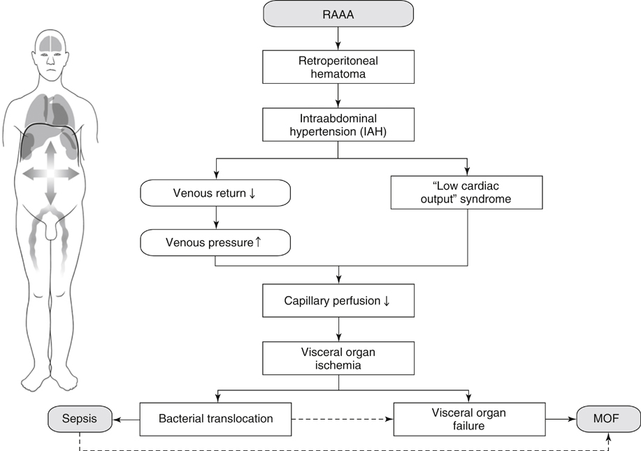
Significant physiologic derangements occur as a result of IAH and affect not only the intraabdominal organs but also every organ system in the body (polycompartment syndrome). All of these pathophysiologic derangements are potentially reversible if IAH is recognized and rapidly treated before significant organ dysfunction has developed. If these derangements are prolonged or untreated, subsequent organ failure occurs at variable pressure thresholds (specific for each organ system) owing to inappropriate tissue perfusion (Figure 1). Preexisting comorbidities, such as chronic kidney failure, pulmonary disease, or cardiovascular disease can further reduce the threshold of IAH that causes the clinical manifestations of ACS.


Stay updated, free articles. Join our Telegram channel

Aug 25, 2016 | Posted by in CARDIOLOGY | Comments Off on Recognition and Treatment of Abdominal Compartment Syndrome Following Ruptured Abdominal Aortic Aneurysm Repair

Full access? Get Clinical Tree

Get Clinical Tree app for offline access