Pulmonary Vasculitis and Hemorrhage

Chapter 59 Pulmonary Vasculitis and Hemorrhage


Pulmonary Vasculitis


Pulmonary vasculitis is defined as inflammation of vessels in the lung of different sizes—pulmonary arteries, veins, and capillaries, as well as bronchial arteries. It usually is only one manifestation of a systemic disorder caused by any of a variety of immunologic mechanisms. Moreover, not all respiratory symptoms occurring in patients with vasculitis are caused by inflammation of pulmonary vessels.


Primary vasculitis is separated from secondary vasculitis. The primary systemic vasculitides are a heterogeneous group of syndromes, of unknown etiology, that share a clinical response to immunosuppressive therapy. Their wide spectrum of frequently overlapping clinical manifestations is defined by the size and location of the affected vessels as well as by the nature of the inflammatory infiltrate. Secondary vasculitis may occur in the context of a well-defined underlying disorder or may be attributable to a specific cause such as collagen vascular disease, infection, or therapeutic or illicit drug use.


Most classification schemes and definitions of the vasculitides are based on the size of the most prominently affected vessels. Definitions, nomenclature, and classification schemes have been and remain subject to change. The terms used in this chapter adhere to the nomenclature and definitions put forth by the Chapel Hill consensus conference in 1992.


Small Vessel Vasculitis


Granulomatosis with Polyangiitis and Microscopic Polyangiitis


Definitions and Nomenclature


GPA is characterized by necrotizing granulomatous inflammation involving the respiratory tract and necrotizing vasculitis affecting small to medium-sized vessels. The most commonly affected vessels are capillaries, venules, arterioles, and arteries, but the wall of the aorta also may rarely be affected by necrotizing granulomatous inflammation—hence the term polyangiitis. Histopathologic documentation of granulomatous involvement of the respiratory tract is not explicitly required. Radiologic evidence or clinical examination findings highly predictive of granulomatous pathology are sufficient. Consequently, the diagnosis of GPA depends on a correlation of clinical, histopathologic, and serologic features. MPA is defined as necrotizing vasculitis with few or no immune deposits, affecting small vessels including capillaries, venules, or arterioles (polyangiitis). Necrotizing arteritis involving small and medium-sized arteries may be present. Necrotizing glomerulonephritis is very common; pulmonary capillaritis resulting in alveolar hemorrhage occurs frequently. The vasculitis of MPA is indistinguishable from that of GPA, and substantial clinical overlap has been observed. For these reasons, the therapeutic approach to patients with GPA and to those with MPA is governed by the same principles, and most clinical studies and therapeutic trials have combined both diseases.


Histopathologic features that define GPA and separate it from MPA include discrete or confluent necrotizing granulomatous inflammation with vasculitis. Fibrinoid necrosis, microabscesses, focal vasculitis, thrombosis, and fibrous obliteration of the vascular lumen also may be seen. Giant cells are a hallmark of the necrotizing granulomatous inflammation of GPA. Atypical and rare histopathologic features of GPA include organizing pneumonia, bronchocentric inflammation, and a marked number of eosinophils in the inflammatory infiltrates.


For treatment stratification, clinical disease activity is categorized as “limited disease” or “severe disease.” Severe disease is either life-threatening or associated with involvement of an organ with potentially irreversible loss of function. Consequently, patients with any of the following disease manifestations should be labeled as having severe disease: alveolar hemorrhage, glomerulonephritis, eye involvement (except mere episcleritis), or nervous system involvement including sensorineural hearing loss. Limited disease includes essentially all patients who have nonsevere disease. The designation limited disease as used in the United States comprises what European investigators have referred to as “early-systemic disease” and “localized disease.” Even though this separation is not based on well-defined biologic distinctions, most disease manifestations leading to the categorization as severe disease are caused by capillaritis. By contrast, most symptoms leading to the classification as limited disease are the result of necrotizing granulomatous inflammation. Patients with limited GPA have a more protracted disease course, a greater likelihood of experiencing a disease relapse after a period of remission, and a higher prevalence of destructive upper respiratory tract disorders (e.g., saddle nose deformity). Tracheobronchial disease involvement also is a feature of GPA not shared with MPA; it may present unique treatment challenges (as discussed later on).


Diagnostic Evaluation


The diagnostic evaluation of patients suspected of having GPA or MPA should include imaging of the chest, pulmonary function testing if the patient has any respiratory symptoms or abnormalities on the chest radiograph, measurements of erythrocyte sedimentation rate, C-reactive protein, complete blood count, serum chemistry panel, urine analysis and microscopy, and testing for ANCA.


Respiratory symptoms usually are associated with unilateral or bilateral radiographic abnormalities including infiltrates, nodules, or mass lesions, which may or may not cavitate. The nodules range in size from a few millimeters to several centimeters across. Solitary nodules also may occur. Unusual manifestations include lymphadenopathy, lobar consolidation, and large pleural effusions.


Tracheobronchial lesions are common and may be asymptomatic or mistaken for evidence of asthma. Pulmonary function testing including inspiratory and expiratory flow-volume loops may provide important clues to the presence of airway narrowing and should be part of the initial evaluation. Bronchoscopic inspection of the tracheobronchial tree is recommended for patients with unexplained respiratory symptoms, abnormalities on pulmonary function tests, or radiographic abnormalities.


Patients with GPA or MPA exhibit variable degrees of elevation of erythrocyte sedimentation rate or C-reactive protein levels. If GPA or MPA is suspected, urine analysis and microscopy and serum creatinine determination are crucial, because early renal involvement may be clinically silent yet can progress rapidly.


In GPA and MPA, ANCA that cause a cytoplasmic immunofluorescence pattern (c-ANCA) on ethanol-fixed neutrophils are caused by the reaction of antibodies with proteinase 3 (PR3) (i.e., PR3-ANCA). By contrast, a variety of antibodies can cause a perinuclear immunofluorescence pattern (p-ANCA) on ethanol-fixed neutrophils. Only those that also react with myeloperoxidase (MPO) are of interest in the context of ANCA-associated vasculitis. Maximal diagnostic accuracy of ANCA testing requires corroboration of a positive target antigen–specific test result (PR3-ANCA or MPO-ANCA) by immunofluorescence assay, or vice versa. Only the PR3-ANCA with c-ANCA combination and the MPO-ANCA with p-ANCA combination are sensitive and specific for ANCA-associated vasculitis.


The clinical utility—as reflected in positive and negative predictive values—of ANCA testing for GPA and MPA is critically dependent on the pretest probability of the disease in the patient tested, as well as on the analytic accuracy of the test method. If applied in patients with clinical features indicating a high pretest probability of GPA or MPA, ANCA testing has a very high positive predictive value. However, occasional false-positive ANCA test results have been reported in a variety of infections. Particularly, subacute bacterial endocarditis may represent a diagnostic dilemma as it may mimic small-vessel vasculitis clinically and has been reported with c-ANCA/PR3-ANCA. With other infections reported with ANCA, either the right pairing of immunofluorescence test results with its corresponding antigen specificity is lacking or their clinical features are distinct from those of GPA or MPA. In patients undergoing evaluation for necrotizing glomerulonephritis with or without alveolar hemorrhage, ANCA may occur in conjunction with anti–glomerular basement membrane (anti-GBM) antibodies. These ANCA usually are of the MPO-ANCA variety. The presence of anti-GBM antibodies seems to determine the prognosis in such double-positive patients.


Most patients with severe GPA or MPA demonstrate a positive result on ANCA testing (for a sensitivity greater than 95%), but up to 30% of patients with limited GPA may not have detectable ANCA. Despite the recognized association between ANCA titers and disease activity, changes in ANCA levels do not reliably predict the disease activity in individual patients. Therefore, serial titers of ANCA should not be used to plan long-term therapy.


Therapy


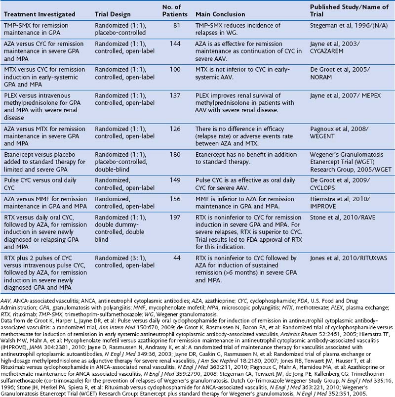
Standard therapy for GPA and MPA currently follows the same basic principles. Methotrexate (MTX) at a dose of up to 25 mg once a week in combination with oral prednisone is considered the standard of care for patients with limited GPA. However, only one prospective randomized trial has compared MTX and cyclophosphamide (CYC) for remission induction in such patients (Table 59-1). The trial, conducted by the European Vasculitis Study Group (EUVAS), showed that MTX is noninferior to CYC for remission induction, but the side effects were less frequent and less severe. The trial also documented that early discontinuation of immunosuppression in patients with ANCA-associated vasculitis is fraught with a high relapse rate. The largest reported group of patients with limited Wegener’s granulomatosis (WG) was treated with MTX for remission induction in the context of the Wegener’s Granulomatosis Trial (WGET). More than 90% of patients achieved remission with this regimen, and more than 70% achieved a sustained remission (lasting longer than 6 months). These rates are equivalent to those achieved with CYC in severe disease, as discussed next.


CYC at a dose of 2 mg/kg/day in combination with prednisone has been the standard of care for patients with severe GPA or MPA until recently. In contrast with the original regimen introduced by Fauci 40 years ago, the current consensus is to limit the duration of CYC therapy to the first 3 to 6 months of remission induction.


Once remission has been induced and the prednisone taper is well under way, CYC should be switched to either azathioprine (AZA), preferred in patients with renal involvement and any degree of renal insufficiency, or MTX. The first option is supported by the results of a randomized trial showing that AZA is as good as CYC for remission maintenance to 18 months. Another randomized controlled trial has shown that MTX and AZA are equivalent for remission maintenance. By contrast, a recent randomized controlled trial that compared mycophenolate mofetil (MMF) with AZA for remission maintenance has shown that MMF is inferior to AZA for this purpose. Thus, the use of MMF for remission maintenance can be supported only for patients who have failed MTX and AZA, or who have contraindications to both agents. The WGET study, in which MTX was used for remission maintenance, confirmed that long-term remission remains an elusive goal for many patients, because remission was maintained in less than half of the patients.


Whenever CYC is used for remission induction, consideration should be given to the patient’s fertility. Young men should be offered sperm banking before therapy is initiated. If time allows, ovarian protection should be offered to young women, in addition to minimizing the cumulative exposure as much as possible.


One randomized controlled trial has evaluated a regimen of intravenous pulse application of CYC compared with daily oral use. The pulse regimen was noninferior to the oral application of CYC for remission, and the frequency of leukopenia, but not infection, was lower. Even though the trial was not powered to detect a difference in relapses, however, the relapse rate was higher after remission induction with the intermittent pulse regimen than with the oral application of CYC.


A metaanalysis of earlier cohort studies had also indicated that intravenous pulse CYC therapy may be safer because of a lower cumulative dose, but a higher relapse rate also was observed after discontinuation. In my own experience, intravenous CYC is best avoided in the intensive care unit setting. However, its use is preferred over oral CYC in patients with questionable compliance, in young women with fertility issues, and in patients who have gastrointestinal problems with oral CYC application.


The four-decade-old standard use of CYC for remission induction in patients with severe GPA and MPA recently has been challenged by the results of two randomized controlled trials. The RAVE (rituximab for ANCA-associated vasculitis) trial was a randomized double-blind, double-placebo-controlled, multicenter trial that compared oral CYC (2 mg/kg/day) to rituximab (RTX) (375 mg/m2 body surface area/week for 4 weeks) for remission induction in severe GPA or MPA in 197 patients. Once remission was achieved between 3 and 6 months, patients randomized to receive CYC were switched to AZA for remission maintenance for 18 months, whereas patients in the original RTX group then received placebo. No difference was observed in rates of achieving remission at the end of 6 months and maintaining remission at 18 months between the two treatment arms. Among the 101 patients who entered the trial with severe relapsing disease, however, RTX proved superior to CYC. The results of the RAVE trial led to approval by the U.S. Food and Drug Administration (FDA) of RTX for remission induction in severe GPA and MPA.


Another randomized controlled open-label trial conducted in 44 patients with newly diagnosed severe ANCA-associated vasculitis with active renal disease, RITUXVAS (Rituximab versus Cyclophosphamide for ANCA-Associated Renal Vasculitis), showed results complementary to those of the RAVE trial. In the RITUXVAS trial, patients were randomized 3 : 1 to receive RTX (together with two pulses of CYC) compared with standard intravenous pulse CYC therapy followed by oral AZA. The primary outcome was sustained remission (of more than 6 months’ duration) at month 12. No difference between the treatment groups was found (RTX 76% versus CYC 82%).


Mycophenolate mofetil (MMF) may represent an alternative to CYC (and RTX) for patients with MPA who have MPO-ANCA and mild renal disease (as defined by creatinine levels less than 3.5 mg/day) and no other life- or organ-threatening disease manifestation. This claim is supported by data from a randomized controlled trial in 35 patients from China comparing oral MMF (1.5 to 2 g/day) with intravenous CYC (0.75 to 1.0 g/m2 once monthly). In addition, all patients received intravenous methylprednisolone bolus therapy (0.5 g/day for 3 days) followed by oral prednisone (0.6 to 0.8 mg/kg for 4 weeks tapered by 5 mg every week to reach 10 mg/day). These regimens demonstrated equivalent efficacy, but MMF was better tolerated than CYC. A prospective pilot trial in 17 patients conducted at the Mayo Clinic achieved similar results.

Stay updated, free articles. Join our Telegram channel

Jun 12, 2016 | Posted by in RESPIRATORY | Comments Off on Pulmonary Vasculitis and Hemorrhage

Full access? Get Clinical Tree

Get Clinical Tree app for offline access