Pulmonary Parasitic Infections


Pulmonary Parasitic Infections


Bobbi S. Pritt, M.D., MSc


Overview

A number of parasites may be found in the human lung, including unicellular protozoa, helminths (worms), and, rarely, arthropods.1,2,3,4,5 The types of parasites may vary significantly with host exposure, age, gender, and immune status.1,2,4,5

Tables 43.1, 43.2, 43.3, 43.4 list the parasites that may infect the lung. The most common protozoan pulmonary diseases worldwide are amebiasis, malaria lung, and toxoplasmosis, while the most common helminthic diseases are dirofilariasis, echinococcosis, paragonimiasis, schistosomiasis, strongyloidiasis, and trichinosis. In addition, three common pulmonary syndromes due to helminths are Loeffler syndrome, tropical pulmonary eosinophilia, and visceral larva migrans.5,6 The anatomic pathologist may play a role in diagnosing all of these infections, although most are usually detected using traditional laboratory methods such as serology and blood film microscopy.


Protozoa

Lung infection by protozoa is usually a component of systemic or disseminated disease. The protozoa most likely to be encountered by the pathologist are Entamoeba histolytica and Toxoplasma gondii. The freeliving amebae (FLA), Acanthamoeba spp. and Balamuthia mandrillaris, are only rarely encountered. It is important to note that protozoal infection is not usually accompanied by peripheral eosinophilia, as is common with many helminth infections.


Entamoeba histolytica (Amebiasis)


General

Entamoeba histolytica is a protozoan parasite that inhabits the human intestinal tract and causes locally invasive and disseminated disease.3,7 Amebiasis is responsible for 40,000 to 100,000 deaths annually, making it the third most common cause of death due to parasitic infection worldwide after malaria and schistosomiasis.8,9 Humans acquire infection primarily through ingestion of environmentally resistant cysts in fecally contaminated food or water. In the United States, disease is seen primarily in immigrants and travelers from endemic areas. Following ingestion of infectious cysts, trophozoites are released into the intestinal lumen and colonize the colon. Most cases of amebiasis (>90%) are asymptomatic, with only a minority of patients developing clinical manifestations.3 When present, symptoms commonly include watery or bloody diarrhea, abdominal pain, and dysentery. In rare cases, trophozoites may enter the portal blood supply and disseminate to other organs.3,10

Pleuropulmonary disease is the second most common form of extraintestinal amebiasis, after the liver.8,11 Nearly all cases result from direct extension of a liver or subphrenic abscess across the diaphragm, and thus, the right lung is most commonly affected. Forms of lung involvement include pleuritis, pleural empyema, pulmonary abscess, bronchohepatic fistula, bronchobiliary fistula, lung abscess, and, rarely, superior vena cava syndrome.8,12 Symptoms may include right upper quadrant pain, pleuritic chest pain, dyspnea, fever, cough, and hemoptysis. Expectoration of thick


red-brown sputum resembling “anchovy paste” or “chocolate sauce” is indicative of a bronchohepatic fistula.8,11,13 Risk factors for pleuropulmonary amebiasis include male sex, atrial septal defect, malnutrition, alcoholism, and immunocompromised state.8,11


TABLE 43.1 Protozoan Parasites of the Human Lung


Parasite/Infection


Infection


Pulmonary Pathology and Manifestations


Geographic Distribution


Acanthamoeba spp. (free-living ameba)


Amebiasisa


Bronchial pneumonia: cough, dyspnea, fatigue, fever


Worldwide, profoundly immunocompromised hosts


Babesia species


Babesiosis


Interstitial pneumonia, acute respiratory distress syndrome: cough, dyspnea, fatigue, fever


Temperate climates worldwide including North America, Europe, and Asia where Ixodes spp. ticks present


Balamuthia mandrillaris (free-living ameba)


Amebiasisa


Bronchial pneumonia: cough, dyspnea, fatigue, fever


Worldwide, usually immunocompromised hosts


Cryptosporidium spp.


Cryptosporidiosis


Bronchitis, interstitial pneumonia: cough, dyspnea, fatigue, fever


Worldwide, usually immunocompromised hosts


Cystoisospora (Isospora) belli


Cyclosporiasis


Interstitial pneumonia: cough, dyspnea, fatigue, fever


Regions of the tropics and subtropics worldwide


Entamoeba histolyticab


Amebiasisa


Abscess, bronchohepatic fistula, liver abscess: cough, hemoptysis, chest pain, right upper quadrant pain


Worldwide, settings with poor sanitation


Leishmania spp.


Visceral leishmaniasis


Interstitial pneumonia, usually a component of disseminated disease: cough, dyspnea, fatigue, fever


Regions of the tropics and subtropics worldwide


Plasmodium falciparumb


Malaria lung


Pulmonary edema, acute respiratory distress syndrome: cough, dyspnea, severe hypoxia, respiratory failure


Regions of the tropics and subtropics worldwide


Trichomonas tenax


Trichomoniasis


Pneumonia: cough, dyspnea, fatigue, fever


Worldwide, associated with poor oral hygiene


Toxoplasma gondiib


Toxoplasmosis


Interstitial pneumonia, bronchiolar exudates, pulmonary edema, cough, dyspnea, tachypnea, +/- fever


Worldwide, profoundly immunocompromised hosts and in neonates


Trypanosoma cruzi


Chagas disease


Interstitial pneumonia, usually a component of disseminated disease: cough, dyspnea, fatigue, +/- fever


Mexico, Central and South America


a The term amebiasis may be used to describe infection with any of the parasitic or free-living amebae but is most commonly used to describe infection with Entamoeba histolytica.
b Most common parasites causing human disease.


Modified from Procop GW, Neafie RC. Human parasitic pulmonary infections. In: Zander DS, Farver CF, eds. Pulmonary Pathology.


Philadelphia, PA: Churchill Livingstone; 2008:287-314; Kim K, Weiss LM, Tanowitz HB. Parasitic infections. In: Broaddus VS, Mason RJ, Ernst JD, et al., eds. Murray and Nadel’s Textbook of Respiratory Medicine. Philadelphia, PA: Elsevier Saunders; 2016:682-698.


TABLE 43.2 Nematode Parasites of the Human Lung


Parasite


Infection


Pulmonary Pathology and Manifestations


Geographic Distribution


Ancylostoma duodenalea


Hookworm infection


Loeffler syndromeb


Tropics and subtropics worldwide with poor sanitation


Ascaris lumbricoidesa


Ascariasis


Loeffler syndrome,b bronchopneumonia due to larvae, rarely adult worms may migrate to trachea, lung, pulmonary artery, and pleurae


Tropics and subtropics worldwide with poor sanitation


Baylisascaris procyonis


Baylisascariasis, visceral larva migrans


Eosinophilic pneumonia: cough, dyspnea, fatigue, fever


North America where raccoons are found


Brugia speciesa


Filariasis


Tropical pulmonary eosinophiliac due to microfilariae, adult worms in pulmonary arteries cause thrombosis and occlusion


Tropics, especially South and Southeast Asia


Dirofilaria immitis and other Dirofilaria speciesa


Dirofilariasis


Pulmonary nodule, lung infarction: cough, hemoptysis, fever, chest pain


Worldwide, including the southeastern United States


Gnathostoma spinigerum


Gnathostomiasis


Bronchitis, pneumonia, cough, hemoptysis, fever, respiratory failure, chest pain, pneumothorax, pleural effusion, expectorated worm


Southeast Asia and Mexico where insufficiently cooked fish, frog, or snake is ingested


Mansonella perstans


Filariasis


Pleural cavity filariasis, microfilariae released into peripheral blood: fever


Parts of Africa, South America


Necator americanusa


Hookworm infection


Loeffler syndromeb


Regions of the tropics and subtropics worldwide with poor sanitation


Strongyloides stercoralisa


Strongyloidiasis, Strongyloides hyperinfection syndrome


Loeffler syndrome,b bronchial infiltration, secondary bacterial pneumonia: cough, dyspnea


Tropics and subtropics worldwide with poor sanitation including southeastern United States (Appalachia)


Trichinella spiralis and other Trichinella species


Trichinosis, trichinellosis


Pneumonia, respiratory failure, usually a component of disseminated disease: cough, dyspnea


Worldwide, where insufficiently cooked flesh of carnivores (pig, boar, bear, walrus) is consumed


Toxocara canis and Toxocara catia


Toxocariasis, visceral larva migrans


Eosinophilic pneumonia; cough, dyspnea, fatigue, fever


Worldwide


Wuchereria bancroftia


Filariasis


Tropical pulmonary eosinophiliac due to microfilariae, adult worms in pulmonary arteries cause thrombosis and occlusion


Tropics, especially South and Southeast Asia


a Most common parasites causing human disease. Other nematodes have rarely been reported to involve the lung. These include anisakiasis (infection with Anisakis spp. and Pseudoterranova decipiens) (eosinophilic pleural effusion); angiostrongyliasis (pulmonary artery thrombosis); capillariasis (Capillaria aerophila) (bronchial infection); pinworm infection (Enterobius vermicularis) (pulmonary nodules, cough, and dyspnea); halicephalobus (Halicephalobus/Micronema gingivalis aka H. deletrix) (associated with fatal encephalitis); lagochilascariasis (Lagochilascaris minor) (pulmonary nodule); and onchocerciasis (Onchocerca volvulus); and syngamosis (Mammomonogamus laryngeus) (cough, hemoptysis, worms attached to mucosa).
b Loeffler syndrome is associated with the pulmonary migration stage of certain intestinal nematodes. It is characterized by self-limited fever, dry cough, wheezing, and peripheral and tissue eosinophilia.
c Tropical pulmonary eosinophilia is a syndrome associated with infection with the filarial worms that cause lymphatic filariasis. It is characterized by chronic low-grade fever, nocturnal paroxysmal dry cough and wheezing, high peripheral and tissue eosinophilia. Microfilariae are rarely seen in tissue sections. When present, they are generally within eosinophilic necrotizing granulomas.


Modified from Procop GW, Neafie RC. Human parasitic pulmonary infections. In: Zander DS, Farver CF, eds. Pulmonary Pathology. Philadelphia, PA: Churchill Livingstone; 2008:287-314; Kim K, Weiss LM, Tanowitz HB. Parasitic infections. In: Broaddus VS, Mason RJ, Ernst JD, et al., eds. Murray and Nadel’s Textbook of Respiratory Medicine. Philadelphia, PA: Elsevier Saunders; 2016:682-698.


TABLE 43.3 Cestodes of the Human Lung


Parasite


Infection


Pulmonary Pathology and Manifestations


Geographic Distribution


Echinococcus granulosusa and other Echinococcus multilocularis


Hydatid cyst; alveolar echinococcosis


Pulmonary cyst; cough, hemoptysis, fever, allergic symptoms, and anaphylaxis with cyst rupture


Pulmonary alveolar echinococcosis is usually spread from the liver


Worldwide, rural sheep-rearing regions and where dogs ingest raw viscera of infected animals


Spirometra spp.


Sparganosis


Pulmonary or pleural nodule, pneumonia: cough, sputum production, fever, chest pain


Worldwide where humans ingest insufficiently cooked frogs and snakes or use frog/snake flesh as a wound poultice


Taenia solium


Cysticercosis


Pulmonary nodules, usually a component of disseminated disease: cough, sputum production


Worldwide where humans ingest insufficiently cooked pork (prerequisite for acquiring the adult tapeworm and shedding infectious eggs into the environment)


a Most common cestode to infect the lung.


TABLE 43.4 Trematodes (Flukes) of the Human Lung


Parasite


Infection


Pulmonary Pathology and Manifestations


Geographic Distribution


Alaria spp.


Alariasis


Single case reported of human respiratory involvement: cough, dyspnea, hemoptysis, fatal asphyxia


Parts of Southeast Asia where insufficiently cooked frogs and snakes are consumed


Clinostomum complanatum


Clinostomiasis


Worm attaches to mucosa of the larynx: throat pain and discomfort, tickling sensation


Parts of Japan and Korea where insufficiently cooked freshwater fish is consumed


Fasciola hepatica


Fascioliasis


Eosinophilic pneumonia, usually associated with liver involvement: cough, dyspnea, fatigue, fever, right hypochondrial pain, chest pain, hepatomegaly


Worldwide, sheep and cattle-rearing regions where raw watercress is ingested


Paragonimus speciesa


Paragonimiasis


Cavitary lesions and pleural effusion: cough, hemoptysis, dyspnea, severe chest pain, night sweats


Parts of Southeast Asia, Africa, North, Central and South America where insufficiently cooked crustaceans are ingested


Schistosoma speciesa


Schistosomiasis, Katayama fever


Pulmonary hypertension, cor pulmonale: dyspnea, fatigue, chest pain, pleural effusion, ascites


Parts of Asia, Africa, South America


a Most frequent trematodes to affect the lung.


Organisms are not usually microscopically detectable in stool of patients with extraintestinal disease; the ova and parasite (O&P) exam is positive in only 35% of cases.8 Therefore, serologic detection of parasite-specific IgG class antibodies is the preferred method of diagnosis.3,7 Microscopic examination of aspirated abscess fluid and sputum and less commonly biopsy of affected organs may also be useful for confirming the diagnosis in conjunction with clinical and radiologic features and serology.7


Radiologic Findings

Chest radiographs and CT scans are commonly used for diagnosing and monitoring extraintestinal amebiasis. Common findings include pleural effusion, pneumothorax, and lung abscess. Pleuropulmonary amebiasis due to extension from a hepatic source is commonly associated with hepatomegaly, elevated right hemidiaphragm, right pleural effusion, and basal lung opacity.8,9,11


Tissue Sampling

Aspirated pleural fluid, abscess material, or expectorated pus generally contains few microscopically recognizable organisms.3,11 In these specimens, detection of E. histolytica DNA using molecular tests such as polymerase chain reaction (PCR) may be useful for confirming the diagnosis. The sensitivity of identifying trophozoites in abscess material may be increased by examining the very last portion of aspirated fluid since this presumably comes from the abscess edge where trophozoites are more prevalent.11 Biopsies and tissue sections should also be taken from the edge of an abscess to increase the likelihood of finding microscopically recognizable trophozoites.


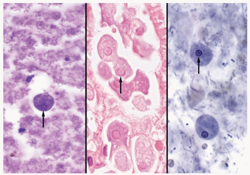
FIGURE 43.1 ▲ In extraintestinal abscesses (left panel, PAS), trophozoites are relatively small with dense cytoplasm and a small nucleus with poorly defined chromatin (arrow). The characteristic peripheral chromatin with central karyosome (nucleolus) may be more readily apparent in invasive intestinal disease (arrow, central panel) but still is not as well defined as in routine stool preparations (arrow, right panel, iron hematoxylin).


Gross and Microscopic Findings

Grossly, amebic abscesses contain yellow-gray to red-brown granular opaque fluid, classically described as resembling “anchovy paste” or “chocolate sauce.”3 Abscesses consist of amorphous necrotic material surrounded by a fibrinous wall and outer rim of edematous host tissue with a mixed inflammatory infiltrate. Neutrophils are generally absent.3 Trophozoites are found most readily in clumps within the fibrin of the abscess wall and can be recognized using standard H&E-stained sections (Fig. 43.1); they generally measure <35 µm in greatest dimension and have dense bubbly cytoplasm that may contain engulfed erythrocytes and a single eccentrically placed round nucleus measuring 4 to 5 µm in diameter.3 Nuclei have characteristic finely clumped peripheral chromatin and a tiny dot-like central karyosome (nucleolus). The trophozoite nucleus may not always be present in each plane of section and the central dot-like karyosome is not as distinct in tissue sections as it is seen in standard stool preparations. Trophozoites may be highlighted using PAS, GMS, and Warthin-Starry stains, while the Brown-Hopps tissue Gram stain may accentuate the nuclear karyosome.3


Treatment and Prognosis

The standard therapy for extraintestinal amebiasis is oral or parenteral metronidazole or other nitroimidazole drug.8,12 As metronidazole will not effectively kill the intestinal cyst forms, patients are also commonly given paromomycin, iodoquinol, or diloxanide furoate to prevent recurrent disease. Surgical resection is contraindicated.8,12 Percutaneous needle or catheter aspiration and drainage of amebic lung abscesses is also not routinely indicated but may be performed in patients who do not improve with metronidazole therapy or who pose a risk for rupture. If untreated, pleuropulmonary amebiasis is usually fatal.


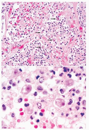
FIGURE 43.2 ▲ Amebic pneumonia due to Acanthamoeba sp./Balamuthia mandrillaris. Low magnification (top) demonstrates multiple trophozoites (arrows) within a neutrophilic alveolar infiltrate. At higher magnification (bottom), characteristic trophozoites with a small round nucleus and large central karyosome can be appreciated.


Toxoplasma gondii (Toxoplasmosis)


General

Toxoplasma gondii commonly causes asymptomatic or self-limited infection, but can also cause life-threatening disease in immunocompromised individuals and devastating congenital infections. As many as one-half of the world’s population is estimated to be infected.5 The prevalence in the United States ranges from 3% to 20%, with the highest prevalence found in individuals living in the central and eastern states and among Native Americans.5

Humans become infected in a variety of manners including ingestion of tissue cysts in undercooked meat, ingestion of oocysts in contaminated food and water, and transplacental transmission from a newly infected mother to her developing fetus.

Immunocompetent individuals are generally asymptomatic but may experience an acute, self-limited mononucleosis-like illness with or without lymphadenopathy, and occasionally accompanied by pneumonia and myocarditis.5 Following primary infection, the parasite remains dormant but may reactivate if the patient becomes immunocompromised. In immunocompromised patients, primary or reactivated infection can result in life-threatening disseminated disease involving the brain and multiple other organs including the lung.14 The brain is the most common site for disseminated disease, but a pulmonary component is seen more than 70% of patients with disseminated disease.5 Congenital infections can also result in pulmonary involvement. The most common signs and symptoms of pulmonary toxoplasmosis are cough, dyspnea, and fever. If untreated, Toxoplasma pneumonia is progressive and fatal.5

Diagnosis of pulmonary toxoplasmosis is usually accomplished by testing for T. gondii-specific IgM and IgG antibodies. False-positive IgM results are not uncommon, and tests for both IgM and IgG antibodies may be negative in profoundly immunocompromised patients.14 Detection of T. gondii DNA in respiratory specimens provides supportive evidence of infection, while detection of tachyzoites in sputa, BAL, or biopsy is diagnostic of acute toxoplasmosis.15


Radiologic Findings

CT scans commonly show diffuse interstitial and alveolar infiltrates.5 Focal consolidation may also be present, while mediastinal and hilar lymphadenopathy are usually absent.

Only gold members can continue reading. Log In or Register to continue

Stay updated, free articles. Join our Telegram channel

Aug 19, 2016 | Posted by in CARDIOLOGY | Comments Off on Pulmonary Parasitic Infections

Full access? Get Clinical Tree

Get Clinical Tree app for offline access