Fig. 17.1
The pulmonary biopsy technique. The biopsy area is grasped with the forceps perpendicular to the pulmonary surface (a). The forceps are closed for 1–2 s and pulled towards the 5-mm insulated trocar. Simultaneously the operator applies a brief current (approximately 1 s) using a foot pedal while pulling the forceps through the distal end of the trocar. The sharp distal tip of the trocar cuts the specimen (b). After this maneuver, the biopsy site blanches and retracts without bleeding or evidence of an air leak (c). A recent experimental study in animal subjects showed that subpleural biopsies obtained during pleuroscopy and deep lung biopsy specimens obtained by electrocautery provided satisfactory material for histologic examination (Emam et al. 2012) (Courtesy C Boutin and Ph Astoul – Marseille – France)
17.3 Indications and Contraindications
17.3.1 Diffuse Lung Disease
In patients with diffuse interstitial lung disease in whom the multidisciplinary integration of clinical and high-resolution computed tomography (HRCT) data, with the addition of BAL and TBLB data, is insufficient to yield a confident diagnosis, current guidelines recommend a surgical lung biopsy in the absence of medical contraindications (Wells and Hirani 2008). A dedicated chest radiologist and chest physician should discuss the HRCT images in advance and decide upon the most appropriate target area to biopsy. There are no randomized controlled trials comparing the three different biopsy techniques (wedge biopsy during VATS, forceps biopsy during VAMT, surgical open lung biopsy). Forceps lung biopsy during VAMT has been used for many years by pulmonologists (Boutin et al. 1982; Loddenkemper and Boutin 1993; Mathur and Loddenkemper 1995). Nevertheless the range of indications for forceps lung biopsies at VAMT in diffuse lung disease has steadily decreased over the last three decades; this has been demonstrated by the change in clinical practice at the Lungenklinik of Berlin where forceps lung biopsy accounted for 22 % of all thoracoscopies in 1971–1979, decreasing to 8 % in 1980–1988, and only 1 % in 1989–1996 (Loddenkemper 1998).
Most studies on VAMT discuss the technical characteristics, diagnostic yield, and potential complications (Boutin et al. 1982; Dijkman et al. 1982), but the diagnostic accuracy of the technique depends on the distribution pattern of the interstitial lung disease, and thus the lobular compartment involved (Fig. 17.2), and the histological specificity of the disease (Vansteenkiste et al. 1999). Only a few papers addressed the issue of biopsy size and histopathological quality (Vansteenkiste et al. 1999; Colt 1995).


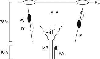
Fig. 17.2
Schematic drawing of the anatomy of the secondary lobule supplied by the bronchovascular bundle with a membranous bronchiole (MB) and a pulmonary artery (PA). The MB branches into the respiratory bronchioles (RB) in the centrilobular region. At the distal end there is the alveolated parenchyma (ALV) with the precapillary arterioles. The lobule is surrounded by the pleura (PL) and the interlobular septum (IS) containing the pulmonary veins (PV) and lymphatics (LY), the latter are also being present in the bronchovascular bundle. During medical thoracoscopy with lung biopsy procedure, the bronchovascular bundle is sampled in 10 % of the specimens and the periphery of the lobule, the IS, and the PL in 78 % of cases (From Vansteenkiste et al. 1999)
A direct comparison of stapled wedge biopsy by VATS to thermocoagulation-assisted forceps biopsy by VAMT has been performed in adult swine with healthy lungs (Colt 1995). Transbronchial biopsy specimens by bronchoscopy are usually <2 mm2 in size, forceps biopsies by VAMT are round-ovoid and range from 2.1 to 6.6 mm in longest diameter or 5–30 mm2 in cross-sectional area, and wedge biopsies by VATS range from 27 to 146 mm in longest diameter (Ayed and Raghunathan 2000). The forceps biopsies contain 300–3,000 readily identifiable alveoli in the specimen, while a wedge biopsy section contains at least 8,000 alveoli. No difference in the number of respiratory bronchioles per mm2 was noted between forceps and wedge biopsy. A greater number of vessels per mm2 were found in wedge biopsies compared to forceps biopsies.
Stay updated, free articles. Join our Telegram channel

Full access? Get Clinical Tree


