4 Problems due to adjustments of the machine settings
To a large extent the quality of an echocardiogram depends on the intrinsic parameters of the probe, but the clinician can still make use of a certain number of adjustments to the echocardiographic apparatus in order to improve the quality of a recording. However, these more or less numerous adjustments vary from one echocardiographic recording to the next. Echocardiography therefore remains an operator-dependent examination. The technical pitfalls resulting from the use of echocardiography concern echocardiographic imaging and Doppler echocardiography, as well as spectral Doppler (pulsed and continuous) and colour Doppler (Box 4.1).
Box 4.1 Pitfalls due to adjustments of the machine settings
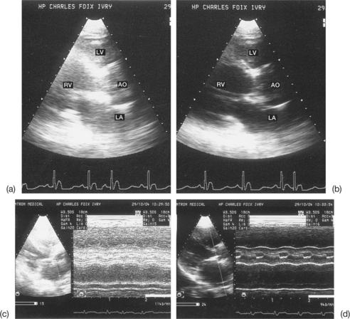
PITFALLS OF ECHOCARDIOGRAPHIC IMAGING (FIG. 4.1)
Inappropriate choice of emission frequency
It is vital to select the optimal emission frequency for the probe generating the ultrasonic waves according to the patient being examined and the diagnostic problem at hand. In general, this frequency falls in the range 2–3.5 MHz in adults and 4–7 MHz in children. A technology known as multifrequency echocardiography, based on the application of variable-frequency ultrasound probes (probes with large bandwidth), enables the operator to select a suitable frequency band for each patient. In fact, the emission frequency chosen is directly responsible for the quality of the image obtained. It is important to remember that the higher the frequency, the better the image resolution. However, at high frequencies the penetration energy of the ultrasonic waves is reduced. This results in an attenuation of the distal echoes, as the deep structures cannot be imaged.
Stay updated, free articles. Join our Telegram channel

Full access? Get Clinical Tree


