Primary and Secondary Vena Cava Tumors


Primary and Secondary Vena Cava Tumors


Thomas C. Bower and Kenneth J. Cherry, Jr.


Primary and secondary tumors of the vena cava are rare and often malignant. They either originate in the vein wall, invade the walls secondarily, or grow within the vena cava as tumor thrombus. They can cause extrinsic compression of the vein lumen. Most patients have advanced local, regional, or distant metastatic disease at the time of diagnosis. Despite advances in adjuvant therapy, survival is measured in months unless surgical resection can be performed. Similar to most cancers and sarcomas, unless a better understanding of the tumor biology is found, development of effective adjuvant therapies will remain problematic.


Tumor Types


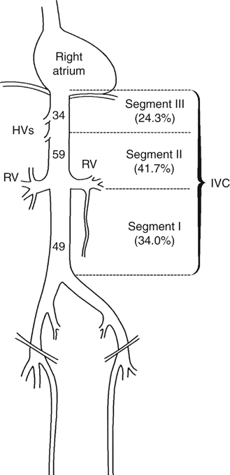
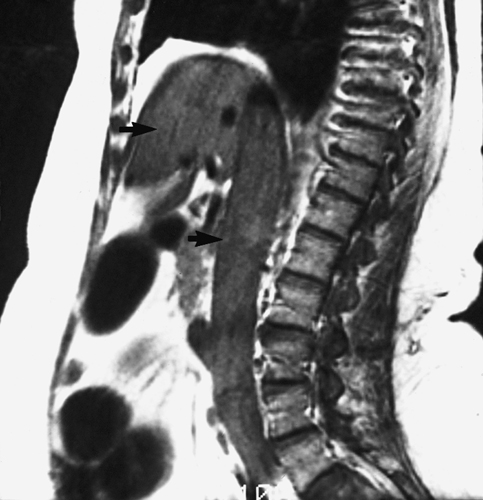
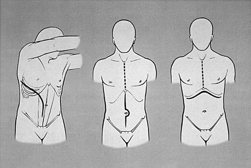
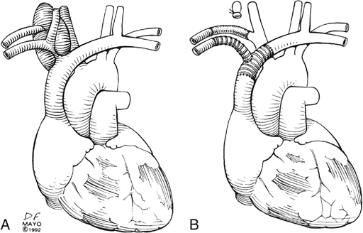
The most common primary tumor of the inferior vena cava (IVC) and superior vena cava (SVC) is leiomyosarcoma. Primary venous leiomyosarcoma involves the IVC in 60% of patients, with three fourths of those involving the suprarenal and infrarenal segments (Figure 1). These tumors are polypoid or nodular, are firmly attached to the vessel wall, and show less intratumor hemorrhage or necrosis than other sarcomas. The most common growth pattern is intraluminal, but the tumor can grow through the vein wall into adjacent structures in advanced cases. Such biologic behavior makes it difficult to differentiate a primary venous leiomyosarcoma from other retroperitoneal sarcomas. Metastatic disease to the lung, liver, kidney, bone, pleura, or chest wall occurs in nearly one half of patients by the time diagnosis is made.


Secondary tumors involve the IVC more often than primary tumors. They can affect any segment of the vena cava, and they often infiltrate its wall (Box 1). Some malignancies exhibit intraluminal tumor thrombus as part of their biologic behavior, with renal cell cancer being the most common. Such renal cell cancers tend to be large (>4.5 cm), and they more often involve the right kidney. Thrombus is isolated to the renal vein–caval confluence in 50%; another 40% have thrombus in the suprarenal IVC, and 10% have thrombus extending into the right heart.


Clinical Presentation


The symptoms and signs of vena cava tumors depend on the level of caval involvement and the type of malignancy. Primary IVC leiomyosarcoma is more common in women than men, occurs over a wide age range, and when symptoms occur, abdominal pain is evident in 66% to 96% of patients. A rare patient with a venous leiomyosarcoma develops a consumption coagulopathy.


SVC obstruction leads to headaches, upper extremity and/or facial swelling or cyanosis, and syncopal events. Patients with SVC obstruction can get markedly short of breath, especially when leaning forward. Cardiac arrhythmias, syncope, pulmonary embolism, pulmonary hypertension with right-sided heart failure, or Budd–Chiari syndrome are seen with suprahepatic caval involvement. Primary or secondary malignancies of the suprarenal IVC rarely cause kidney failure because of retrograde outflow through the tributaries of the left renal vein, the lumbar veins, and the paravertebral venous plexus. Abdominal pain and a palpable mass are seen with malignancies at any level in the abdomen. Lower extremity edema, neuropathy, and dilated abdominal wall veins are late signs of infrarenal IVC obstruction.


Evaluation


The Mayo Clinic has a multidisciplinary team to evaluate and treat patients with venous tumors. Specialists include medical or surgical oncologists and vascular, hepatobiliary, urologic, orthopedic, neurologic, and cardiothoracic surgeons as needed. The goals of evaluation are to determine the type and extent of the malignancy, to search for distant metastatic disease, and to assess the severity of venous obstruction. Computed tomography (CT), magnetic resonance imaging (MRI), and ultrasonography (US) are most often used to accomplish these goals.


If a tumor proves to be localized and there is no evidence of metastatic disease, preoperative risk assessment is done, which includes cardiopulmonary assessment. Patient performance status influences the decision to operate. Those who are bedridden or need significant help in daily care are not candidates for operation. CT and MRI are the two most useful studies because assessment of local extent of the tumor is done by axial, coronal, and sagittal images. Venous phase imaging has all but eliminated venacavography as a diagnostic study, unless intraluminal biopsy is needed. The deep veins of the lower extremities are imaged by US to exclude occult bland DVT. MRI is particularly useful in defining the extent of intracaval tumor thrombus, and transesophageal echocardiography is added when there is concern of thrombus extension into the right heart (Figure 2).


Treatment


Operation is offered to a select group of patients with localized disease, good performance status, and satisfactory cardiopulmonary function. The operative treatment and approach depends on the type of tumor and the extent of caval involvement, the degree of caval obstruction, the status of the collateral veins; and the patient’s body habitus. Tumors of the SVC are approached through a median sternotomy, with the excision extended toward either side of the neck as needed to complete the resection and to perform venous reconstruction. Malignancies involving the suprahepatic or suprarenal IVC, which require concomitant liver resection, are approached through an 8th or 9th space right thoracoabdominal excision, through a bilateral subcostal incision if the costal flare is wide, or, less often, through a midline abdominal incision that is extended into the chest through a median sternotomy as needed.


In patients with renal cell cancer and intracardiac tumor thrombus the approach is through a median sternotomy and midline abdominal incision, though occasionally the sternotomy is combined with a bilateral subcostal incision. The infrarenal IVC is best approached through a midline incision, which allows access to the common iliac veins for inflow control (Figure 3).


Most patients with malignant involvement of the SVC are treated by stenting, but for the rare patient in whom tumor-free margins are anticipated, the SVC can be replaced using either femoral vein, spiral saphenous vein, or synthetic graft. For the latter, we prefer externally supported expanded polytetrafluoroethylene (ePTFE) grafts (Figure 4). Should the tracheal or esophageal wall require resection, femoral vein is a good alternative.


Stay updated, free articles. Join our Telegram channel

Aug 25, 2016 | Posted by in CARDIOLOGY | Comments Off on Primary and Secondary Vena Cava Tumors

Full access? Get Clinical Tree

Get Clinical Tree app for offline access