Acute inferior myocardial infarction is characterized infarction of inferior wall of left ventricle that is territory of right or circumflex coronary artery, frequently occurs with abrupt obstruction of any else of these arteries. We report a case of inferior myocardial infarction that diagnosed clinically and electrocardiographically but angiographically no any culprit lesion in right or circumflex coronary artery but serious lesion and thrombus without complete obstruction in proximal left anterior descending artery.
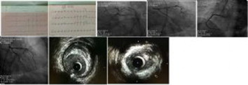
A 38 years old man without previous cardiac history and risk factories is admitted to our coronary care unit with typical chest pain last as 20 minutes as in last 24 hours. The patient feels chest discomfort during moderate exercise last a month. Physical examination revealed blood pressure and heart rate were 117/70mmHg and 80/beat per minute. Heart sounds were normal with no any murmur. Respiratory sounds were normal. His ECG revealed sinüs rhythm, ST segment elevation and pathological Q wave in lead 2,3 and aVF and posterior leads (figure1). Echocardiographic findings included left ventricular ejection fraction %60, left ventricular apical hypokinesia and left ventricular diastolic dysfunction (grade 1).
Laboratory findings revealed maximum troponin I:0,684 ng/ml, maximum CKMB:54U/l and other routine biochemical parameters were normal. The patient was taken to catheter laboratory for coronary angiography and PCI. In coronary angiography, right and circumflex coronary artery were normal but there is serious stenosis and thrombus formation in the proximal of left anterior decsending artery(figure2). We decided to perform intravascular ultrasonography to evaluate the degree of lesion severity. Intravascular ultrasonography showed serious stenosis and thrombus (figure3). The patient was referred to cardiovascular surgery for coronary artery bypass grafting.
Acute STEMI is a condition required emergency revascularization. Our case confirms clinically acute inferior myocardial infarction. We performed coronary angiography but we did not see any culprit lesion in right or circumflex coronary artery and their branches. However, there is severly stenosis and hazy view in proximal segment of left anterior descending artery. Because of inflamatory markers are normal, miyocarditis is low probability. Both echocardiographically left ventricle apical hypokinesia and seriously stenosis and thrombus (see figure 2-3) in the proximal segment of LAD was associated acute coronary syndrome. We referred the patients to cardiovascular surgery for CABG.


