Figure 49.1
Detection of a parenchymal fistula. To localize the site of a pulmonary air leak, the pleural cavity is filled with saline solution. The anesthesiologist ventilates the atelectatic lung, and the surgeon holds it underwater; the air leak is identified by following the bubbles rising in the water. The bronchial stump must always be checked separately to rule out the existence of a bronchopleural fistula. This underwater procedure is repeated after any closure of a pulmonary fistula to make sure the fistula sealing was successful and to rule out the existence of other lesions
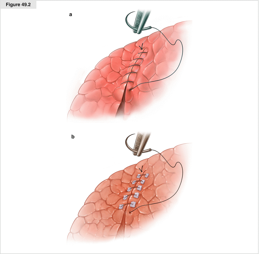
Figure 49.2
Closure of parenchymal fistula by suturing. (a) After the origin of the fistula is identified, the leakage site of the pulmonary parenchyma is closed by overstitching with nonabsorbable sutures, such as propylene 3.0 or 4.0. If there is only a small lesion caused by a ruptured bleb, a single U-shaped suture may be enough to achieve airtightness. (b) In patients who previously underwent wedge resection or interlobar division of the pulmonary parenchyma during a lobectomy, the cause of a prolonged air leak is often a broken row of sutures over a distance of several centimeters. In these cases, we sew uninterrupted, nonabsorbable sutures in two layers along the leakage. For reinforcement and to avoid further damage to vulnerable lung tissue, Teflon pledgets may be placed on both sides of the parenchyma where suturing is planned. The sutures are then sewed deeply through the lung parenchyma surrounding the fistula and through the Teflon pledgets, which also serve as an abutment for a safe knot
Bronchial Fistula Following Right Upper Lobectomy
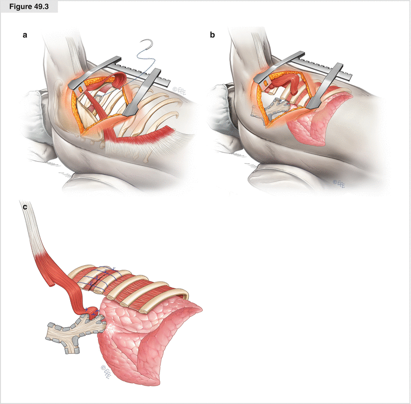
Figure 49.3
Secondary suturing of the open stump and coverage by a pedicled flap of latissimus dorsi muscle. (a) Reopening of the chest with direct access to the empyema cavity is performed best by posterolateral thoracotomy on the right side to avoid disturbance of the potentially adherent residual lung. If the wall of the unhealed bronchial stump is vital and well-supplied with blood, the bronchus is resutured with four to six interrupted 3.0 polypropylene sutures. (b) For further protection of the still vulnerable, resutured stump, coverage of the stump with more vital tissue is recommended. In this location, a pedicled flap of latissimus dorsi muscle is well-suited for bronchial stump coverage. Therefore, the muscle is separated widely from the subcutaneous fat and the slips of the serratus anterior muscle in the depth of the chest wall. The thoracodorsal neurovascular bundle must be treated carefully to guarantee a blood supply to the flap. The pedicle is localized 2–3 cm medial to the anterior muscle border and about 9 cm below the axillary apex. Creation of the pedicled flap is completed by ligation of the muscle at its lumbar attachment. Before the muscle flap is transposed, the neighboring rib close to the pedicle is partially resected to provide the flap with an unobstructed entrance into the pleural cavity. The size of the gap must fit the diameter of the pedicle to make sure there is no compression that may disturb the blood supply. (c) The free end of the flap is transferred through the costal gap to the bronchial stump without being twisted. There, it is tacked to the surrounding tissue so that the stump is fully covered by the flap. Ideally, the muscle flap not only covers the bronchial stump, but also, at least partially, fills and thus minimizes the infected cavity of empyema
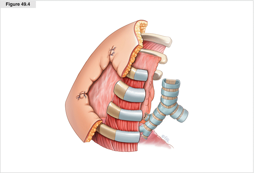
Figure 49.4
Open thoracic window. An alternative method for managing an open bronchial stump following right upper lobectomy with consecutive pleural empyema is to create a small open-window thoracostomy in the right axillary region. To do this, an axillary thoracotomy is performed. The subcutaneous tissue and axillary fat are divided so that the tissue can be used later to cover the edges of the thoracostomy. When preparation reaches the ribs, the second and third rib are exposed from all sides by removing the intercostal muscles and periosteum. These two ribs then are resected over a few centimeters, depending on the patient’s height. The entrance created into the pleural cavity must be wide enough to provide access to the whole empyema cavity, including the open bronchial stump. To fix the thoracostomy, the skin-covered, subcutaneous, and fatty tissue flaps are wrapped around the stumps of the resected ribs and the intact neighboring ribs, where they are fixed by uninterrupted, nonabsorbable, outside-inward sutures placed around the stable surrounding ribs. Following diligent debridement, the cavity is plugged with moist surgical drapes, and the thoracostomy is covered with a sterile dressing. After the procedure, the dressing is changed once or twice a day in the patient’s room. As soon as the pleural cavity has been definitely purified, if the wound remains clean and the patient’s general condition is good enough to undergo another surgical intervention, closure of the open thoracic window by thoracoplasty may be discussed
Bronchial Fistula Following Left Upper Lobectomy
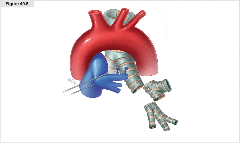
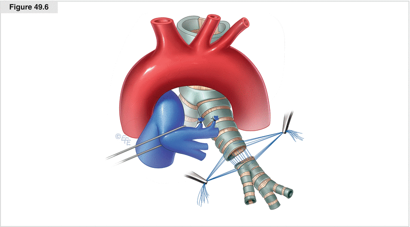
Figures 49.5 and 49.6
Secondary bronchial sleeve resection. To avoid loss of healthy lung tissue, sleeve resection of the open bronchial stump may be performed. The anatomic conditions for secondary sleeve resection are often favorable after lobectomy of the left upper lobe. After (redo) thoracotomy on the left side, the left mainstem bronchus is mobilized to confirm that the anatomy will allow a secondary sleeve resection. The pulmonary artery, located close to the bronchus, is mobilized first and retracted off the bronchus anteriorly. To gain enough room centrally, the ligamentum arteriosum must be divided. When the left mainstem bronchus and the lower bronchus of the left lung have been freed, both are divided by scalpel to allow sleeve resection of the leaking bronchus. The cut is made in healthy bronchial areas with sufficient blood supply to the cutting edges to avoid problems with the healing of the anastomosis. The surgeon must ensure that the superior segmental bronchus of the lower lobe is not injured and there is enough bronchial tissue left to create a tension-free anastomosis. The two resulting bronchial ends are approximated. The anastomosis is started with an uninterrupted polydioxanone suture (e.g., PDS 4.0) of the dorsal part. This suture is fixed by tying it with corner seams stitched on each side of the suture row. The remaining cartilaginous portion of the bronchi is closed by interrupted PDS sutures. The stitches are placed at a 3-mm distance in a way that compensates for the difference in caliber of the bronchi. For further protection of the bronchial suture, a pericardial fat pad based on a central pedicle is prepared and wrapped around the anastomosis, where it is fixed by two or three sutures (see Chap. 18)
Bronchial Fistula Following Left or Right Lower Lobectomy
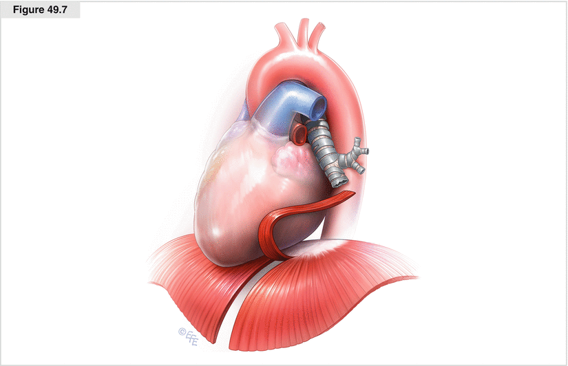
Figure 49.7


Creation of a pedicled diaphragmatic flap. Because of the proximity of the bronchial stump after lower lobectomy to the diaphragm, the use of a pedunculated diaphragmatic flap is an appropriate measure to seal the leak in the bronchial stump in case of a bronchial fistula after lobectomy of the right or left lower lobe. After (redo) thoracotomy, the diaphragm is checked to make sure it provides enough tissue to cover the leaking stump and to verify that the distance is not too great to be bridged by the flap. To create the pedunculated flap with a width of approximately 2 cm, two parallel longitudinal incisions are made in the diaphragm, following the course of the muscle fibers from front to back. The dorsal end of the resulting strip is cut between two clamps, and the cutting edges are closed with ligatures
Stay updated, free articles. Join our Telegram channel

Full access? Get Clinical Tree


