Figure 40.1
Positioning and approach. After the induction of general anesthesia, a double-lumen endotracheal tube is used to facilitate single-lung ventilation. Because blood loss is often significant with this procedure, an arterial line and central venous line also are used to aid in arterial and venous pressure monitoring. The patient is placed in the lateral decubitus position. A posterolateral thoracotomy incision is extended downward toward the costal margin over the sixth rib and posteriorly beyond the tip of the scapula. A more limited incision, such as a standard posterolateral thoracotomy incision, may be used to start the procedure. After intrathoracic assessment of resectability, the incision may be extended anteriorly as mentioned above. The incision is made downward over the sixth rib, and the periosteum is lifted off the rib superiorly and inferiorly (inset). A periosteal elevator is used to separate the rib from the surrounding soft tissue. A rib cutter is used to separate the rib anteriorly and posteriorly, then the rib is removed. Dissection between the parietal pleura and endothoracic fascia is begun
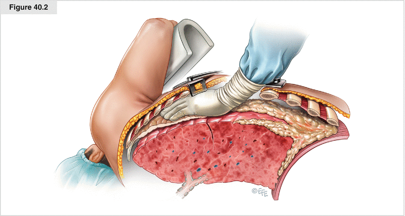
Figure 40.2
Dissection of the extrapleural plane. The plane then takes on a cephalad direction toward the apex from the posterolateral direction. Both blunt and sharp dissections are used, with sharp dissection saved mainly for areas of dense adhesions. The apex is often relatively free of tumor compared with the diaphragmatic surface. Dissection in this direction first makes exposure and inferior dissection easier. The placement of two Finochietto retractors, one anteriorly and the other posteriorly, greatly enhances the visualization of the thoracic cavity. As the dissection moves anteriorly, care must be taken not to avulse the internal mammary vessels off the subclavian vessels. Blunt dissection with sponge sticks can effectively separate the pleura from the anterior chest wall close to the mammary vessels, but sharp dissection is recommended once the vessels are found. As the area of dissection increases, previous areas of dissection are packed with laparotomy pads for hemostasis
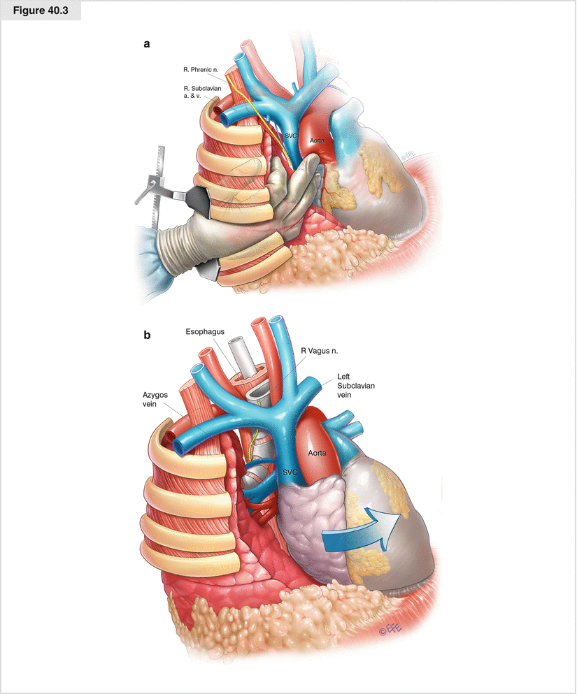
Figure 40.3
Dissecting the apex. The subclavian vessels sit at the apex of the thoracic cavity. As the dissection progresses, great care must be used in dissection around the subclavian vessels at the apex. As the dissection proceeds toward the mediastinum, the azygous vein and superior vena cava (SVC) are approached with great caution. Once the upper portion of the lung is completely mobilized from the chest wall, the superior and posterior hilar structures are disclosed. The aorta and arch vessels must be identified and dissected carefully (a). Injury to the phrenic and recurrent laryngeal nerves also must be avoided. After the right upper lobe and right mainstem bronchus are seen, dissection of the esophagus is begun (b). A nasogastric tube helps in palpating and identifying the esophagus. The SVC is then gently dissected off the specimen, and the dissection continues to the posterior aspect of the pericardium. The vagus and phrenic nerves are identified again and protected. a. artery, n. nerve, v. vein
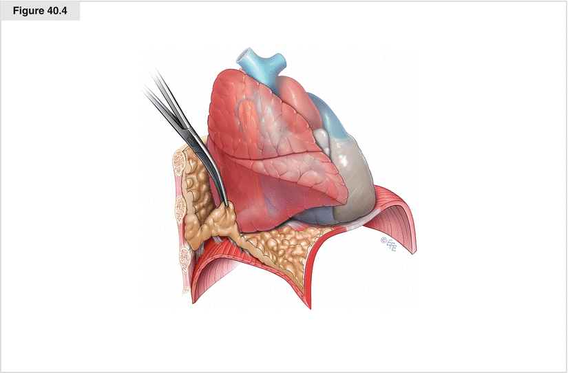
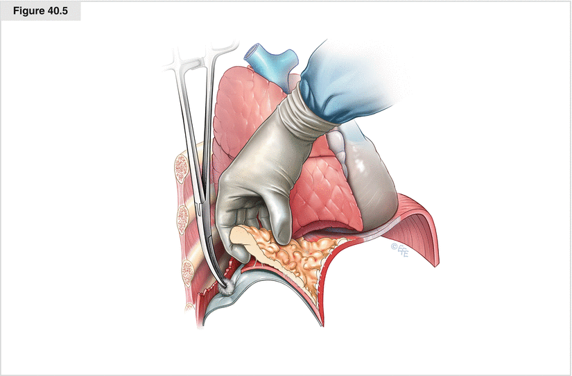
Figure 40.4
Elevation of the parietal pleura off the diaphragm. The extent of tumor at the level of the diaphragm determines whether resection is required. If the parietal pleura peels off the diaphragm easily, the diaphragm can remain intact. If necessary, the entire diaphragm may be resected and reconstructed with Gore-Tex (W. L. Gore & Associates, Elkton, MD)