Pericardial Cyst
Presentation
A 77-year-old man with a past medical history significant for hypertension presents to his primary care physician with complaints of left-sided chest pain. The patient denies cough, hemoptysis, fever, chills, night sweats, anorexia, and weight loss. He is a nonsmoker. He reports a previous episode of left-sided chest pain, about 10 years ago, at which time he was diagnosed with a chest x-ray abnormality that has been followed with serial computed tomography (CT) scans and no recommended intervention.
▪ Chest X-rays
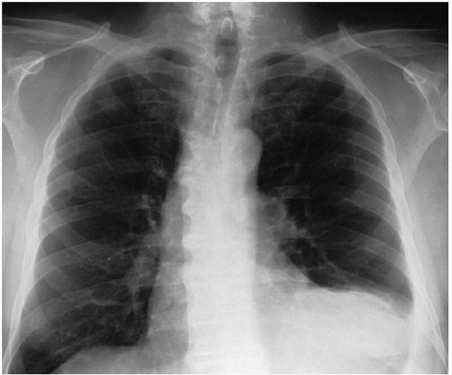 Figure 52-1 |
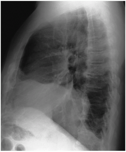 Figure 52-2 |
▪ CT Scans
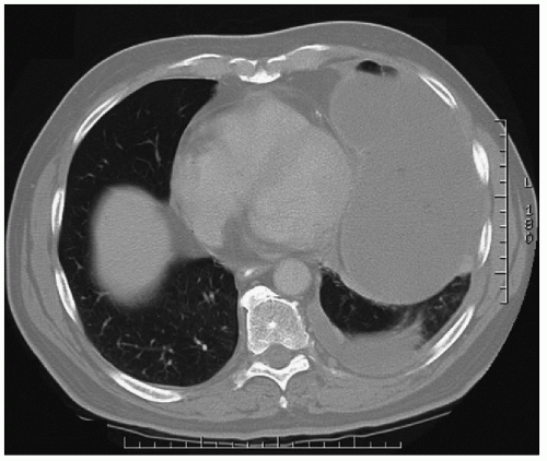 Figure 52-3 |
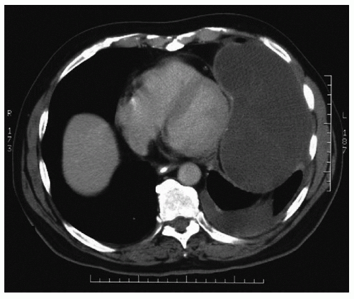 Figure 52-4 |
CT Scan Report
There is a loculated fluid collection in the left lower thorax measuring 15 cm × 8 cm. There is a small to moderate amount of free-flowing left-sided pleural fluid. On the CT scan performed 10 years earlier, the cyst measured 5 cm and was associated with minimal atelectasis of the left lower lobe. Serial CT scans are evaluated that demonstrate a gradual increase in size. The last scan, performed 4 years before the current study, measured the cyst at 16.5 cm, which was associated with minimal atelectasis.
Stay updated, free articles. Join our Telegram channel

Full access? Get Clinical Tree


