Percutaneous Catheter-based Therapy for Valvular Heart Disease

40 Percutaneous Catheter-based Therapy for Valvular Heart Disease


Charles Dotter is credited with noting that the stenotic severity of a high-grade iliac lesion was lessened when a diagnostic catheter was passed through it. Early vascular efforts used progressively larger catheters to open the lesions by blunt dilatation. Eventually, this approach using progressively larger bougies was replaced by the use of elastic balloon-tipped catheters, first for peripheral vascular disease, then for coronary angioplasty. Reports from the National Heart, Lung, and Blood Institute (NHLBI) Registry, the Mansfield Balloon Catheter Registry, and large institutional experiences subsequently shaped the development of percutaneous balloon procedures for stenotic valvular lesions. Percutaneous valvuloplasty (or valvotomy) has now become the standard of care for the treatment of certain patients with congenital pulmonary valve and aortic valve stenosis and for a subset with rheumatic mitral stenosis. Recently, percutaneous valve replacement or repair has been proposed as a feasible advance in percutaneous therapies applicable to both stenotic and regurgitant valve lesions. It is anticipated that selected patients will be candidates for these novel approaches in the near future. This chapter is meant to provide an overview of all of these percutaneous therapeutic approaches to valvular heart disease.


Pulmonary Valve Stenosis


Pathophysiology


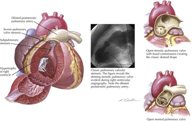
Pulmonary valve stenosis results from fusion of the valve cusps during mid- to late fetal development. The most common form of isolated right ventricular (RV) obstruction, pulmonary valvular stenosis, occurs in approximately 7% of individuals with congenital heart disease (see also Chapters 50 and 52). Pulmonary valve stenosis may be associated with significant RV hypertrophy and infundibular narrowing. The fusion of the valvular cusps produces a classic systolic “doming” appearance angiographically (Fig. 40-1). Tissue pads within the valve sinuses may exist and result in a thickened, rigid valve that is considered dysplastic (a common finding in Noonan’s syndrome). Excessive thickening in dysplastic valves renders the valve unsuitable for percutaneous valvuloplasty, although attempts have occasionally been successful. Similarly, narrowing of the RV outflow tract limits the efficacy of percutaneous balloon techniques. Acquired forms of stenosis are rare (i.e., carcinoid).


Percutaneous Balloon Pulmonary Valvuloplasty


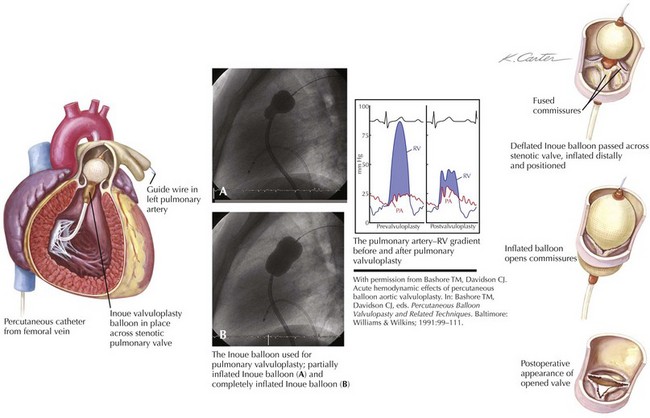
Figure 40-2 demonstrates the gradient between the right ventricle and the pulmonary artery (PA) before and after successful percutaneous balloon valvuloplasty. The RV outflow tract may have considerable muscular subpulmonic stenosis, which may be masked when valvular obstruction is present. The sudden removal of the valvular stenosis after the procedure may result in acute decompensation from marked RV infundibular obstruction, sometimes called the “suicide RV.” Fluid loading, calcium channel blockers, and β-blockers can be used for emergent treatment. After pulmonary valvuloplasty, the subpulmonic hypertrophy may regress considerably over the next several months.


Technique


Before the procedure, RV angiography in the cranial right anterior oblique and straight lateral views is performed. Pulmonary angiography assesses preprocedural pulmonic regurgitation. Severe pulmonic regurgitation (3+ or more) is a contraindication to valvuloplasty; severe regurgitation as a result of the procedure represents an adverse outcome. Baseline annular size is determined by echocardiography, MRI, or contrast angiography. In the cardiac catheterization laboratory, a catheter (with radiopaque markers a known distance apart) may be used for angiography at the valve level to determine appropriate balloon size. Quantitative angiographic methods may be similarly applied.


The dilating balloon or balloons are percutaneously inserted into the femoral vein without a sheath. The maximum inflation of the balloon(s) should be equal to 1.2 to 1.4 times the estimated annular size (see Fig. 40-2). In contrast to the aortic valve (see “Aortic Valve Stenosis”), the pulmonic valve is elastic and often requires oversizing for adequate results. The goal of the procedure is a final peak-to-peak valvular gradient less than 30 mm Hg by cardiac catheterization. Recurrence rates are much lower if that threshold is reached. A single balloon, often 23 mm in diameter in adults, may be used, although two balloons side by side may be necessary in patients with a large annulus. In some laboratories, trefoil or bifoil balloon catheters are available and preferred. The Inoue mitral valvuloplasty balloon (Toray Industries, Inc., Tokyo, Japan) has increasingly been used for pulmonary valvuloplasty because of its stability during inflation.


Careful measurement of postprocedural gradients allows differentiation of infundibular stenosis from residual valvular stenosis. Postprocedural PA angiography evaluates the severity of pulmonic regurgitation as a result of the procedure, while postprocedural RV angiography addresses the presence and significance of infundibular stenosis.


Percutaneous Pulmonary Valve Replacement


In 1992, the first percutaneous catheter-mounted pulmonary heart valve procedure using a stented porcine bioprosthetic valve was implanted in an animal model. In 2000, Bonhoeffer and colleagues were the first to treat a human with a percutaneous pulmonary valve implanting of a stent-mounted bovine jugular vein valve in an RV-to-PA conduit of a 12-year-old. The remarkable success of the index case led quickly to its application in eight additional cases, with major hemodynamic improvement in both stenosis and regurgitation seen in five of the eight. The procedure has now been applied to a wider group of patients with available follow-up at a median of 3 years. The overall results remain encouraging.


Aortic Valve Stenosis


Pathophysiology


The normal aortic valve has thin, flexible cusps composed of three tissue layers sandwiched between layers of endothelium on both sides of the valve. The layers include a fibrosa with collagen fibers oriented parallel to the leaflet that support the major leaflet, a ventricularis layer composed of elastic fibers oriented perpendicularly to the leaflet edge that provide flexibility, and a spongiosa layer of loose connective tissue in the basal third of each leaflet.


Congenitally deformed aortic valves have fused commissures and can generally be described as either unicuspid or bicuspid. Unicuspid valves are inherently stenotic at birth and cause symptoms early in life. Unicuspid aortic valves account for approximately 10% of all cases of isolated aortic valve stenosis in adulthood, whereas bicuspid aortic valves account for approximately 60% of isolated aortic valve stenosis in patients aged 15 to 65 years of age. Bicuspid aortic valves generally have two cusps of nearly equal size with a false commissure (raphe). Over time, progressive valvular fibrosis and calcium deposition occur, worsening the functional stenosis. Some commissural fusion between the functioning leaflets may occur, but the major limitation is often valvular rigidity from calcium buildup and scarring. High serum lipids may contribute to the degeneration of these valves similarly to those patients with calcific aortic stenosis.


Aortic valve stenosis in elderly persons generally involves a trileaflet valve and probably represents a continuum, from benign aortic valvular sclerosis to severe aortic valvular stenosis. The prevalence of aortic valve sclerosis has been reported to be 25% in individuals older than 65 years of age, with severe aortic valve stenosis evident in 1% to 2% of the population. There is growing evidence that the mechanism of calcific aortic valve stenosis in the elderly is related to atherosclerosis and not to what has commonly been referred to as a “degenerative” process. Little commissural fusion exists; large accretions of calcium can be present in the sinuses of Valsalva. The leaflets gradually lose their flexibility as a result of these calcium deposits. In calcific aortic valve stenosis, the minimal reduction in the gradient that can be obtained by balloon procedures has generally been attributed to cracks in the calcific nodules, cuspal tears, and aortic wall expansion (Fig. 40-3).


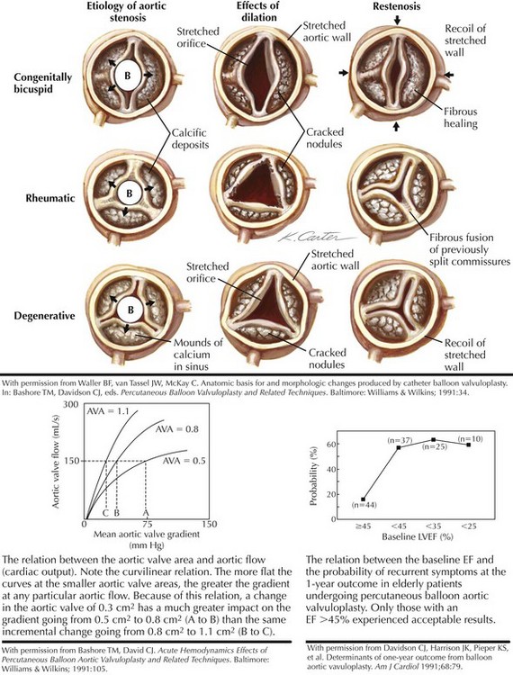
When left ventricular (LV) outflow is obstructed at the valvular level, a gradient develops between the left ventricle and the aorta (see Fig. 40-3). The relationship between the gradient and the aortic valve area (AVA) is complex, however, and depends on the severity of the lesion as measured by the AVA and on the cardiac output or the aortic flow. After aortic valvuloplasty, aortic flow may increase because of an improvement in the cardiac output or the development of aortic regurgitation. Either result could increase the gradient, even if the actual AVA also increases. Alternatively, the cardiac output may fall, and the gradient may appear lower even if the AVA has increased. Thus, the short-term postprocedural valvular gradient change may not always reflect the actual change in the AVA.


Using just the change in the AVA can also be problematic for other reasons. For instance, if the baseline AVA is severe, an improvement in the AVA of 0.3 cm2 from baseline has a dramatic effect on the peak LV systolic pressure (e.g., when the AVA increases from 0.5 to 0.8 cm2), but if the baseline AVA is less severe, the same incremental change may have much less consequence (e.g., when the AVA increases from 0.8 to 1.1 cm2). Hence, either an improvement in the gradient or an improvement in both the gradient and the final valve area can be used to define a successful result (i.e., a final valve gradient of <50 mm Hg, a 50% improvement in the AVA, or both).


Indications for Intervention


The decision whether to intervene in aortic valve stenosis usually depends on the presence of symptoms of congestion, angina, or exertional syncope and an assessment of the likelihood of improvement in AVA. Serial measurements of transvalvular pressure gradients by Doppler echocardiography can be helpful. When the maximum Doppler velocity exceeds 4 m/s (estimated gradient of 64 mm Hg), symptoms emerge relatively quickly. A change in the Doppler gradient of more than 0.3 m/s within 1 year also portends symptoms. Thus, recent guidelines suggest that severe aortic stenosis is present when the Doppler estimated peak instantaneous gradient is greater than 64 mm Hg, the mean gradient is greater than 40 mm Hg and/or the estimated AVA is less than 1.0 cm2. Because of the variable means for measuring valvular gradients and the dependence of the valve gradient on the aortic valvular flow and the effective orifice area, the use of a specific AVA to make a decision on the need for an operation is always tenuous. This may be particularly true in patients with a low cardiac output and low gradient, but severe aortic stenosis by calculated AVA. In this situation, the use of an inotropic agent or nitroprusside to augment aortic flow may help determine whether the low output (and the subsequently low gradient) is a consequence of the valvular stenosis or is attributable to poor ventricular function. A very high brain natriuretic protein level may suggest a poor prognosis in this group, even if aortic valve replacement (AVR) is performed.


In neonates and very young children, the initial success rates for percutaneous intervention are not encouraging, although older children and young adults may benefit and should be considered for the procedure as a temporizing measure. In older adults, surgical intervention has consistently proven superior to percutaneous balloon valvuloplasty. The use of percutaneous balloon valvuloplasty in adults with either bicuspid or calcific aortic stenosis should be restricted to situations in which the risk of surgical intervention is very high (e.g., in a pregnant patient or in an elderly patient with cardiogenic shock), because of the generally poor results. In these circumstances, percutaneous balloon valvuloplasty may serve as a bridge to eventual AVR. Also, in the rare elderly adult with preserved LV systolic function and severe aortic valve stenosis who is not a candidate for surgical AVR because of comorbid conditions, valvuloplasty can provide short-term symptomatic benefit or be used as a bridge to eventual AVR.


Technique of Percutaneous Balloon Aortic Valvuloplasty


In contrast to pulmonary valvuloplasty, the balloon catheter used for aortic valvuloplasty should have a maximum inflated diameter slightly smaller than the measured size of the aortic annulus. In adults, a 20-mm-diameter balloon is usually used, although a 23-mm balloon may be required for larger patients. Brief, rapid RV pacing during positioning and inflation of the aortic balloon across the stenotic valve lowers cardiac output transiently and allows for more stable positioning. The balloon catheter is placed in the middle of the valve plane and manually inflated, using dilute (25%) radiographic contrast in saline (Fig. 40-4). Inflation pressures do not seem to influence the outcome significantly, and these pressures are no longer measured. Usually one to three separate 15- to 20-second inflations are adequate.
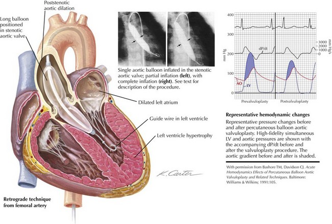


Whether the approach is percutaneous (via the femoral artery, with or without a sheath), cut-down (using the brachial artery), or transseptal (using an antegrade approach to the aortic valve via the right femoral vein), similar results are obtained. The transseptal approach is particularly useful in patients with significant aorto-iliac atherosclerosis, a common problem in elderly individuals. Following the transseptal puncture, a 0.038-inch wire is navigated through the left atrium and the left ventricle, across the aortic valve, and down the descending aorta for stability. The intra-atrial septum is predilated using an 8-mm balloon catheter before insertion of the aortic valvuloplasty balloon catheter. The remainder of the procedure is similar to the retrograde approach.


Acute Results and Complications


The mean aortic gradient can be expected to fall from about 55 to 29 mm Hg acutely, with the AVA increasing from 0.5 to 0.8 cm2 with no measurable change in cardiac output.


In those patients for whom pressure-volume data were derived before and immediately after the procedure, systolic function was largely unchanged, with the ejection fraction (EF) rising only slightly, the peak positive dP/dt falling slightly, and stroke volume and peak and end-systolic wall stress all modestly reduced. A negative impact was noted acutely on diastolic measures of ventricular function, including a significant decrease in peak negative dP/dt and a prolongation of tau (a measure of active diastolic relaxation). Transient mild ischemia during the procedure was considered responsible for some of the acute changes.


Results in children and neonates vary broadly depending on the patient’s clinical status and associated cardiac anomalies. Many neonates with critical aortic valve stenosis have severe LV hypoplasia or endocardial fibroelastosis and do poorly with either percutaneous aortic valvuloplasty or surgery. After the neonatal period, the results from valvuloplasty improve. Data from 232 patients with a mean age of approximately 9 years showed the aortic gradients decreased approximately 60% from about 75 mm Hg to 30 mm Hg after percutaneous balloon valvuloplasty. The procedure seems often to work reasonably well in the adolescent age group, offering an important opportunity to delay surgery until the individual has reached full adult size. It should be noted that, even with an excellent initial outcome, restenosis will occur over time.


The rate of serious life-threatening complications from aortic valvuloplasty is remarkably low given the elderly population in whom it is usually applied. Almost all protocols for calcific aortic stenosis require patients to be noncandidates for surgical intervention. In a review of 791 such patients, in-hospital mortality rates were 5.4% with a risk of serious morbidity (cerebrovascular accident, cardiac perforation, myocardial infarction, or serious aortic regurgitation) of up to 1.5%. Vascular complications were overwhelmingly the greatest complicating feature, with a 10.6% incidence. The common practice today of using vascular occlusion devices after the procedure has virtually eliminated major vascular injury as a concern.


In the NHLBI Registry of 671 patients, complications were considerable. At least one complication was reported in 25% of the patients within 24 hours, and 31% had some complication before hospital discharge. The most common complication was the need for transfusion (23%), followed by the need for vascular surgery (7%), cerebrovascular accident (3%), systemic embolization (2%), or myocardial infarction (2%). All-cause mortality was 3%, with death usually related to multiorgan failure and poor preprocedural LV function. In patients who survived to 30 days, 75% had improved by at least one NYHA functional class.


Jun 12, 2016 | Posted by in CARDIOLOGY | Comments Off on Percutaneous Catheter-based Therapy for Valvular Heart Disease

Full access? Get Clinical Tree

Get Clinical Tree app for offline access