and Alessandra Cancellieri2
(1)
Department of Radiology, Bellaria Hospital, Bologna, Italy
(2)
Department of Pathology, Maggiore Hospital, Bologna, Italy
Radiology | Giorgia Dalpiaz |
Pathology | Alessandra Cancellieri |
Cystic pattern | Definition | Page 204 |
Cystic signs | Thin-walled cysts or not-walled cysts Thick-walled cysts Cysts with irregular shape Cysts with regular shape Bunch of grapes sign Incidental lung cysts | Page 205 Page 206 Page 207 Page 207 Page 208 Page 209 |
Table of diseases | Page 209 |
Cystic Pattern
Definition
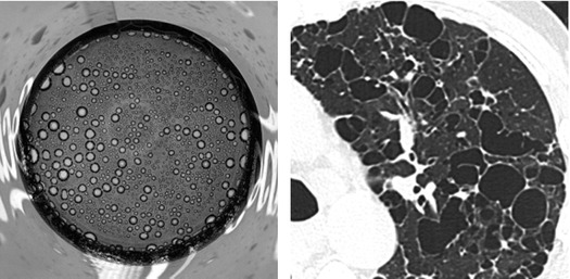
Cystic pattern is present when multiple roundish, well-defined, air-containing spaces (“holes”) are variably scattered throughout the pulmonary parenchyma. The “holes” appear black on HRCT and white on pathological specimens.
These “holes in the lung” may be due to dilatation of bronchial structures, abnormal distension of alveolar spaces, focal destruction of lung parenchyma, or late stage of even cavitation of solid lesions. In elderly patients, lung cysts have also been reported in asymptomatic nonsmoking individuals, raising the possibility that they may represent part of the aging process.
Holes in the lung

The cystic pattern should not be confused with the dark lung pattern. In both models, the elementary lesions are hypodense on HRCT, but, in the cystic pattern, these lesions are focal and not diffuse and their density is that of pure air containing units, as black as the ambient air outside the chest or inside the trachea.
Sometimes, sporadic cysts may be present as ancillary signs in patients with other main signs belonging to other patterns, e.g., subacute hypersensitivity pneumonitis (HP) and desquamative interstitial pneumonia (DIP) (please refer to chapter “Alveolar Pattern”).
The possible HRCT signs are:
Thin-walled or not-walled cysts
Thick-walled cysts
Cysts with irregular shape
Cysts with regular shape
Bunch of grapes sign
Incidental lung cyst
The prevalent distribution of the cysts together with the presence of nonparenchymal signs may be helpful for the diagnosis of a specific disease (please see the Table at the end of this Chapter).
Gupta N (2015) Diffuse cystic lung disease. Part I. Am J Respir Crit Care Med 191(12):1354
Gupta N (2015) Diffuse cystic lung disease. Part II. Am J Respir Crit Care Med 192(1):17
Raoof S (2016). Cystic Lung Diseases: Algorithmic Approach. Chest;150(4):945
Cystic Signs
Thin-Walled Cysts or Not-Walled Cysts
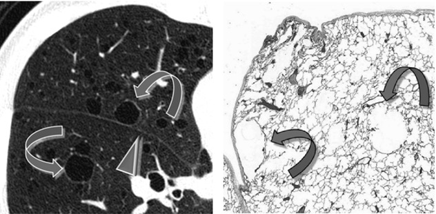
On HRCT thin-walled cysts ( ) present a white encircling rim of a thickness similar to the fissure (►). These types of cysts are often secondary to check-valve mechanisms in the context of a normal parenchyma (e.g., LAM) (
 ).
).
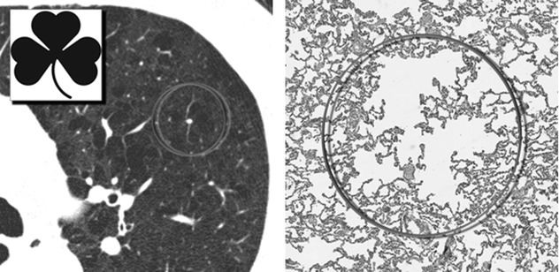
) present a white encircling rim of a thickness similar to the fissure (►). These types of cysts are often secondary to check-valve mechanisms in the context of a normal parenchyma (e.g., LAM) ().Not-walled cysts appear as roundish black holes with invisible walls, surrounded by normal lung parenchyma. Sometimes a central nodular or branching opacity representing the centrilobular artery is seen ( ). From time to time this type of black hole may present a clover aspect (
 ). These types of cysts often are the result of local destruction of lung parenchyma (e.g., emphysema) (
 ).
).
). From time to time this type of black hole may present a clover aspect (). These types of cysts often are the result of local destruction of lung parenchyma (e.g., emphysema) ().Bubble-like cysts, cysts with thin (or no) wall
Diseases having cysts with thin (or no) wall:
Emphysema, centrilobular (images above ): it is the prototype of cystic diseases without wall; however, it may display thin-walled cysts and, occasionally, it can hardly be differentiated from LAM. In these cases, the upper lobe predominance is a key for the differential diagnosis. Paraseptal emphysema is often visible also in the subpleural space in single layer. The anamnesis of smoking habit is also crucial for the diagnosis.
Lymphangioleiomyomatosis (LAM) (images above ): the disease typically involves young women in reproductive age, and the cysts are characteristically thin-walled and round in shape (“lacy” appearance). The most useful sign to differentiate LAM from LCH is the distribution of the cysts. Unlike in LCH, cysts are diffused throughout the lungs and they may involve the juxtaphrenic recesses.
Langerhans Cell Histiocytosis (LCH), end–stage disease: in this phase of disease the cysts may present a thin wall similarly to LAM; however, they are bizarre in shape and predominate in the lung apices, with relative sparing of the lung bases. Crucial is also the anamnesis of smoking habits.
Stay updated, free articles. Join our Telegram channel

Full access? Get Clinical Tree


