and Alessandra Cancellieri2
(1)
Department of Radiology, Bellaria Hospital, Bologna, Italy
(2)
Department of Pathology, Maggiore Hospital, Bologna, Italy
Radiology | Giorgia Dalpiaz | |
Pathology | Alessandra Cancellieri |

Septal pattern | Definition | Page 50 |
Septal signs | Septal thickening Subpleural Interstitial thickening Peribronchovascular thickening | Page 51 Page 51 Page 52 |
Subset smooth and table | Page 53 | |
Subset nodular and table | Page 54 |
Septal Pattern
Definition
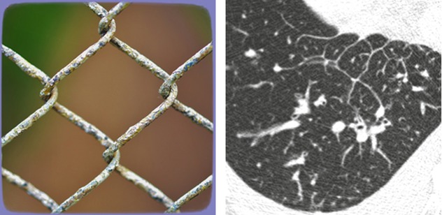
A septal pattern is present when thickening of the perilobular interstitium and bronchovascular bundle, both in the centrilobular core and at the central level, is visible. The final effect is that of a regular network of white lines. Lobular architecture is preserved. Septal pattern can be smooth or nodular in contour depending on the different pathological processes.
The septal pattern may be due to the filling of the interstitium by fluid, neoplastic cells, or inflammation.
Reticular pattern with preserved architecture, regular linear pattern


Reticular pattern may be also “irregular” due to fibrosis, and therefore this subtype is included and explained in the fibrosing pattern. In this case, the architecture is not preserved resulting in a distorted net.
The signs of septal pattern are:
Septal thickening
Subpleural interstitial thickening
Peribronchovascular thickening
The prevalent distribution of the signs together with the presence of non-parenchymal signs may be helpful for the diagnosis of a specific disease (please see also the tables at the end of this chapter).
As well as in septal diseases, in which this pattern is predominant, there are other diseases in which septal pattern (reticular pattern with preserved architecture) may be found, albeit less important or sporadic. They are therefore described in the relevant chapter (e.g., associated with ground-glass opacity in diffuse alveolar hemorrhage, pneumonia, and NSIP or associated with nodules and/or cysts in LIP).
Andreu J (2004) Septal thickening: HRCT findings and differential diagnosis. Curr Probl Diagn Radiol 33(5):226
Hansell DM (2008) Fleischner Society: glossary of terms for thoracic imaging. Radiology 246(3):697
Webb WR (2006) Thin-section CT of the secondary pulmonary lobule: anatomy and the image – the 2004 Fleischner lecture. Radiology 239(2):322
Septal Signs
Septal Thickening
White lines 1–2 cm in length outlining the polygonal boundaries of secondary lobules. A few lines inside the lobule may be also visible. Lobules delineated by thickened septa (➨) commonly contain a visible dot-like or branching centrilobular pulmonary artery (▶).
Stay updated, free articles. Join our Telegram channel

Full access? Get Clinical Tree


