Pathophysiology of Venous Varicosities and Venous Ulcers


Pathophysiology of Venous Varicosities and Venous Ulcers


S. Martindale, M. Chung and P.J. Pappas


Chronic venous insufficiency (CVI) is the seventh leading cause of chronic debilitating disease in the United States. An estimated 25 million people in the United States have varicose veins, 2 million to 6 million have more advanced forms of CVI (swelling, skin changes), and nearly 500,000 have painful venous ulcers. The chronicity of this disorder and the lack of effective therapies place a heavy economic burden on the health care system.


Macroscopic Alterations


The venous system consists of thin-walled, low-pressure conduits. Skeletal muscle contractions in the upper and lower extremities propel blood forward, and a series of intraluminal valves prevents retrograde flow or reflux. Venous hypertension secondary to valvular incompetence is the inciting event that results in the biochemical, microscopic, and macroscopic changes that ultimately manifest as symptoms such as lower extremity edema, pain, itching, skin discoloration, varicose veins, and, in the severest form, venous ulceration. These clinical signs and symptoms collectively refer to chronic venous disorders; manifestations specific to abnormal venous function are known as CVI.


Valvular incompetence is the most common sequela to symptomatic deep vein thrombosis (DVT). It develops in 20% to 50% of patients who have had DVT, depending on the extent of the initial thrombosis. The remaining 50% to 80% have no identifiable cause and are designated as having primary reflux. Patients with primary reflux are hypothesized to have intrinsic or genetic vein wall abnormalities that predispose to varix formation.


Whatever the initiating event, several unique anatomic and biochemical abnormalities have been observed. Normal and varicose greater saphenous veins (GSVs) are characterized by a collagen matrix that provides strength, elastic fibers that provide compliance, and three distinct muscle layers within their walls. The media contains an inner longitudinal and an outer circular layer, and the adventitia contains a loosely organized outer longitudinal layer.


In normal GSVs, these muscle layers are composed of smooth muscle cells (SMCs) that appear spindle shaped (contractile phenotype) when examined with electron microscopy. The cells lie in close proximity to one another, are in parallel arrays, and are surrounded by bundles of regularly arranged collagen fibers.


In varicose veins, the orderly appearance of the muscle layers of the media is replaced by an intense and disorganized deposition of collagen. Collagen deposits separate the normally closely opposed SMCs and are particularly striking in the media. SMCs appear elliptical, rather than spindle shaped, and demonstrate numerous collagen-containing vacuoles that impart a secretory phenotype. Increased phosphorylation of the retinoblastoma protein, an intracellular regulator of cellular proliferation and differentiation, has been observed in varicose veins and might contribute to this process. Similarly, in vitro co-culture studies of SMCs and endothelial cells (ECs) demonstrate SMCs with a contractile phenotype. The addition of an endothelin-1 antagonist to this SMC–EC co-culture system caused the SMCs to change from a contractile to a secretory phenotype. These two observations suggest that endothelin-1 and the retinoblastoma protein might modulate SMC differentiation and contribute to the formation of varicose veins.


Biochemical and functional analyses of varicose veins demonstrate alterations in collagen, elastin, and endothelin content as well as contractility abnormalities. Tissue cultures have demonstrated that smooth muscle cells derived from varicose veins show increased expression and synthesis of type I collagen and decreased synthesis of type III collagen and fibronectin. Gandhi and colleagues quantitatively demonstrated an increase in collagen content and a decrease in elastin content compared to normal GSVs. The net increase in the collagen-to-elastin ratio suggested an imbalance in connective tissue matrix regulation. Lowell’s group evaluated the contractile responses of varicose and normal GSV rings to noradrenaline, potassium chloride, endothelin, calcium ionophore A23187, and nitric oxide. This study demonstrated decreased contractility of varicose veins when stimulated by noradrenaline, endothelin, and potassium chloride. Similarly, endothelium-dependent and -independent relaxations after A23l87 or nitric oxide administration were diminished compared to normal GSV relaxation.


The mechanisms responsible for decreased varicose vein contractility appear to be receptor mediated. Decreased endothelin B receptors have been observed in varicose veins compared to normal GSVs. Feedback inhibition of receptor production secondary to increased endothelin-1 is postulated to mediate the decreased receptor content in varicose vein walls. This hypothesis correlates with in vitro co-culture experiments. Lack of endothelin-1 receptors or endothelin-1 blockade with receptor antagonists both result in decreased contractility.


Microscopic Alterations


Numerous theories regarding the etiology of venous stasis ulceration have been proposed. However, attention is currently focused on white cell activation. This leukocyte-trapping theory states that venous hypertension causes the less deformable white blood cells to be trapped in the venous microcirculation, where they migrate into the dermal interstitium and cause damage by releasing toxic metabolites. The first step in the process is extravasation of macromolecules (fibrinogen and α2-macroglobulin) and red blood cells (RBCs) into the dermal interstitium secondary to prolonged venous hypertension.


RBC degradation products and interstitial protein extravasation are potent chemoattractants and presumably represent the initial underlying chronic inflammatory signal responsible for leukocyte recruitment. It has been assumed that these cytochemical events are responsible for the increased expression of intercellular adhesion molecule-1 (ICAM-1) on endothelial cells of microcirculatory exchange vessels observed in CVI dermal biopsies. ICAM-1 is the activation-dependent adhesion molecule used by macrophages and lymphocytes for diapedesis. Both these cells have been observed by immunohistochemistry in the interstitium of dermal biopsies. However, a morphometric assessment of the dermal microcirculation has identified macrophages and mast cells only and has questioned the role of lymphocytes in CVI dermal pathology.


Extracellular Matrix Alterations


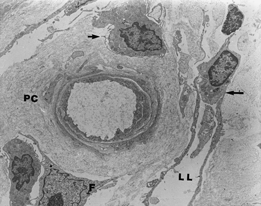
Once leukocytes migrate to the extracellular space, they localize around capillaries and postcapillary venules. The perivascular space is surrounded by extracellular matrix (ECM) proteins. Adjacent to this perivascular cuff and throughout the dermal interstitium is an intense and disorganized collagen deposition. This perivascular cuff and the accompanying collagen deposition are the sine qua non of the dermal microcirculation in CVI patients (Figure 1). The cuff is a ring of ECM proteins consisting of collagens type I and III, fibronectin, vitronectin, laminin, and tenascin. The cuff was once thought to be a barrier to oxygen and nutrient diffusion. Evidence now suggests, however, that cuff formation is an attempt to maintain vascular architecture in response to increased mechanical load.


Stay updated, free articles. Join our Telegram channel

Aug 25, 2016 | Posted by in CARDIOLOGY | Comments Off on Pathophysiology of Venous Varicosities and Venous Ulcers

Full access? Get Clinical Tree

Get Clinical Tree app for offline access