Fig. 2.1
Pathophysiological determinants of GERD. GERD is very heterogeneous in presentation encompassing strictly mucosal consequences, typical reflux symptoms, atypical reflux symptoms, and hypersensitivity syndromes. The one thing that all manifestations have in common is in being triggered by reflux events, that being the overarching definition of the disease in the Montreal scheme
Although GERD is widely reported to be one of the most prevalent clinical conditions afflicting the gastrointestinal tract, incidence and prevalence figures must be tempered with the realization that there has been no “gold standard” on which to base these figures. Thus, epidemiological estimates regarding GERD make assumptions, the most obvious being that heartburn is a symptom of GERD and that when heartburn achieves a certain threshold of frequency or severity, it defines GERD. Applying that methodology, weekly heartburn has a prevalence estimated at 24 % among 18–24-year-olds and 33 % in those over 55 years of age [2]. With respect to esophagitis, early reports using ambulatory esophageal pH monitoring to define GERD found that 48–79 % of patients with pathologic acid exposure had esophagitis [3]. More recently, a population-based study found endoscopic esophagitis in 22 % of 226 individuals with heartburn at least once weekly [2]. GERD is equally prevalent among men and women, but there is a male preponderance of esophagitis (2:1 to 3:1) and of Barrett’s metaplasia (10:1) [4]. Pregnancy is strongly associated with GERD such that 48–79 % of pregnant women complain of heartburn [5]. All forms of GERD affect Caucasians more frequently than other races [2].
A provocative epidemiological observation was of the striking inverse time trends in the prevalence of GERD- and H. pylori-related peptic ulcer disease [6]. Furthermore, GERD patients with esophagitis are less likely to have H. pylori infection and H. pylori infection is associated with a decreased prevalence of Barrett’s metaplasia and esophageal adenocarcinoma [7]. Thus, epidemiological data suggest a complex relationship between H. pylori and GERD, which is to some degree dependent of the associated pattern of gastritis. If the dominant H. pylori strains within a population primarily result in corpus-dominant gastritis as in Japan [8], the prevalence of GERD in that population will be lower than it would be in the absence of H. pylori infection.
Mechanisms of Reflux
Under normal conditions, reflux of gastric juice into the distal esophagus is prevented by the esophagogastric junction (EGJ), which is an anatomically complex area whose functional integrity has been attributed to a multitude of mechanisms. Quite possibly each functional component is operant under specific conditions, and the global function of the EGJ as an antireflux barrier is dependent on the sum of the parts. The greater the dysfunction of the individual mechanisms of competence, the worse the overall antireflux integrity of the EGJ. By extension, the greater the degree of EGJ incompetence, the worse the severity of GERD.
Functional Constituents of the Esophagogastric Junction
Conceptualized as an impediment to reflux, the EGJ is generally viewed as an anatomically complex high-pressure zone at the distal end of the esophagus that isolates the esophagus from the stomach. The esophagus traverses the diaphragmatic hiatus and joins the stomach in a nearly tangential fashion. Thus, there are several potential contributors to EGJ competence, each with unique considerations: the lower esophageal sphincter (LES), the influence of the diaphragmatic hiatus, and the muscular architecture of the gastric cardia that constitutes the distal aspect of the EGJ.
The LES is a short segment of tonically contracted smooth muscle at the distal end of the esophagus. Resting LES tone varies among normal individuals from 10 to 30 mmHg relative to intragastric pressure, and continuous pressure monitoring reveals considerable temporal variation. Large fluctuations of LES pressure occur with the migrating motor complex; during phase III, LES pressure may exceed 80 mmHg. Lesser fluctuations occur throughout the day with pressure decreasing in the postcibal state and increasing during sleep [9]. The genesis of LES tone is a property of both the smooth muscle itself and of its innervation. Vagal afferents as well as both vagal and sympathetic efferents modulate LES pressure [10]. Efferent innervation is mediated through myenteric plexus neurons that can effect either LES contraction or relaxation. Synapses between the efferent vagal fibers and the myenteric plexus are cholinergic. The postganglionic transmitter effecting contraction is acetylcholine, while NO is the dominant inhibitory transmitter with VIP serving some type of modifying role [11]. Hence, at any given moment, LES pressure is affected by myogenic factors, intra-abdominal pressure, gastric distention, peptides, hormones, various foods, and many medications (Table 2.1).
Table 2.1
Factor affecting LES pressure
Increase LES pressure | Decrease LES pressure | |
|---|---|---|
Foods | Protein | Fat |
Chocolate | ||
Ethanol | ||
Peppermint | ||
Hormones | Gastrin | Secretin |
Motilin | Cholecystokinin | |
Substance P | Glucagon | |
Gastric inhibitory polypeptide | ||
Vasoactive intestinal polypeptide | ||
Progesterone | ||
Neural agents | Alpha-adrenergic agonists | Alpha-adrenergic antagonists |
Beta-adrenergic antagonists | Beta-adrenergic agonists | |
Cholinergic agonists | Cholinergic antagonists | |
Serotonin | ||
Medications | Metoclopramide | Nitrates |
Domperidone | Calcium channel blockers | |
Prostaglandin F2α | Theophylline | |
Cisapride | Morphine | |
Meperidine | ||
Diazepam | ||
Barbiturates |
Physiological studies suggest that the EGJ extends distal to the squamocolumnar junction implying that structures in the proximal stomach are involved [12]. Anatomical studies attribute this distal portion of the EGJ to the opposing sling and clasp fibers of the middle muscle layer of gastric cardia [13, 14]. In this region, the lateral wall of the esophagus meets the medial aspect of the dome of the stomach at an acute angle, defined as the angle of His. Viewed intraluminally, this region extends within the gastric lumen, appearing as a fold that has been conceptually referred to as a flap valve because increased intragastric pressure would force the fold against the medial wall of the stomach, sealing off the entry to the esophagus [15, 16]. Of note, this distal aspect of the EGJ is particularly vulnerable to disruption as a consequence of anatomical changes at the hiatus because its entire mechanism of action is predicated on maintaining its native geometry.
Surrounding the LES at the level of the SCJ is the diaphragmatic hiatus, most commonly comprised of the right diaphragmatic crus. Two flattened muscle bundles arising from the upper lumbar vertebra incline forward to arch around the esophagus, first diverging like a scissor and then merging anterior with about a centimeter of muscle separating the anterior rim of the hiatus from the central tendon of the diaphragm. The hiatus is a teardrop-shaped canal and is about 2 cm along its major axis. Recent physiological investigations have advanced the “two-sphincter hypothesis” for maintenance of EGJ competence, suggesting that both the LES and the surrounding crural diaphragm serve a sphincteric function. Independent control of the crural diaphragm can be demonstrated during esophageal distention, vomiting, and belching when electrical activity in the crural diaphragm is selectively inhibited despite continued respiration [17, 18]. This reflex inhibition of crural activity is eliminated with vagotomy. On the other hand, crural diaphragmatic contraction is augmented during abdominal compression, straining, or coughing [19]. Additional evidence of the sphincteric function of the hiatus comes from manometric recordings in patients after distal esophagectomy [20]. These patients still exhibited an EGJ pressure of about 6 mmHg within the hiatal canal despite having sustained surgical removal of the smooth muscle LES.
Mechanisms of EGJ Incompetence in GERD
The dominant mechanism protecting against reflux varies with physiological circumstance. For example, the intra-abdominal segment of the LES may be important in preventing reflux associated with swallowing, the crural diaphragmatic may be of cardinal importance with abrupt increases in intra-abdominal pressure, and basal LES pressure may be of primary importance during restful recumbency. As any of these protective mechanisms are compromised, the deleterious effect is additive resulting in an increasing number of reflux events and consequently increasingly abnormal esophageal acid exposure.
Investigations have focused on three dominant mechanisms of EGJ incompetence: (1) transient LES relaxations (TLESRs) without necessary anatomical derangement; (2) LES hypotension, again independent of anatomical abnormality; or (3) anatomical distortion of the EGJ inclusive of (but not limited to) hiatus hernia. Which reflux mechanism dominates seems to depend upon a number of factors. While TLESRs typically account for up to 90 % of reflux events in normal subjects or GERD patients without hiatus hernia, patients with hiatus hernia have a more heterogeneous mechanistic profile with reflux episodes frequently occurring in the context of low LES pressure, straining, and swallow-associated LES relaxation [21]. Further complicating the issue, prolonged ambulatory high-resolution manometry studies demonstrate that patient often flips back and forth between a hernia and non-hernia configuration of EGJ pressure morphology [22]. These observations suggest that the integrity of the EGJ is dependent on both the LES and the diaphragmatic hiatus. In essence, with normal anatomy, gastroesophageal reflux requires a “two-hit phenomenon” to the EGJ. Inhibition of both the LES and crural diaphragm is required for reflux to occur; physiologically this occurs only in the setting of a TLESR. In contrast, patients with hiatal hernia may exhibit preexisting compromise of the hiatal sphincter. In that setting, reflux can occur with only relaxation of the LES, as may occur during periods of LES hypotension or even deglutitive relaxation.
Transient Lower Esophageal Sphincter Relaxations
There is compelling evidence that TLESRs are the most frequent mechanism for reflux during periods of normal LES pressure (>10 mmHg). TLESRs occur independently of swallowing, are not accompanied by peristalsis, are accompanied by diaphragmatic inhibition, and persist for longer periods than do swallow-induced LES relaxations (>10 s) [23]. Of note, prolonged manometric recordings have not consistently demonstrated an increased frequency of TLESRs in GERD patients compared to controls [24]. However, the frequency of acid reflux (as opposed to gas reflux) during TLESRs has been consistently reported to be greater in GERD patients [25].
Recognizing the importance of TLESRs in promoting reflux, investigators have studied it intensively. The stimulus for TLESRs is distention of the proximal stomach, not surprising given that transient LES relaxation is the physiological mechanism for belching [26]. TLESR can be experimentally elicited by either gaseous distention of the stomach or distention of the proximal stomach with a barostat bag. Furthermore, the degree to which TLESR frequency is augmented by gastric distention is directly related to the size of hiatus hernia suggesting that the associated anatomical alteration affects the afferent mechanoreceptors responsible for eliciting this reflex [27]. The most likely candidate for the afferent receptor is the intraganglionic lamellar ending or IGLE [28]. Intraganglionic lamellar endings are found at the receptor end of vagal afferents innervating the gastric cardia and are activated by applied tension [29]. The frequency of TLESRs is also greater in an upright posture. The vagal afferents from the gastric cardia then project to the nucleus tractus solitarii in the brain stem and subsequently to the dorsal motor nuclei of the vagus. Finally, dorsal motor nucleus neurons project to inhibitory neurons localized within the myenteric plexus of the distal esophagus. Furthermore, TLESR is an integrated motor response involving not only LES relaxation but also crural diaphragmatic inhibition, costal diaphragm contraction, esophageal longitudinal muscle contraction, gastric fundus contraction, and contraction of the rectus muscles of the abdominal wall [23, 30, 31]. The TLESR reflex is abolished by vagotomy [32]. Recently, animal and human experiments have demonstrated that TLESRs can be inhibited by gamma-aminobutyric acid receptor type B agonists (such as baclofen) and mGluR5 antagonists, suggesting a potential alternative approach to the treatment of GERD [33].
Lower Esophageal Sphincter Hypotension
Although a hypotensive LES predisposes to reflux, this is actually a rarely observed mechanism in patients without hiatus hernia [21]. In fact, free reflux is observed only when the LES pressure is within 0–4 mmHg of intragastric pressure. More commonly, strain-induced reflux occurs when a hypotensive LES is overcome and blown open in association with an abrupt increase of intra-abdominal pressure. However, strain-induced reflux is mechanistically unlikely with normal function of crural diaphragm, which reflexively contracts in association with abdominal straining. The crural diaphragm, however, is progressively less functional with increasing size of hiatal hernia as demonstrated in a study of strain-induced reflux that modeled the interaction between the LES and hiatal hernia concluding that there was an important interaction between the two [34].
Another important consideration is that only a minority of patients with gastroesophageal reflux disease have a hypotensive fasting LES pressure (usually defined as <10 mmHg) [46]. This observation can be reconciled somewhat when one considers the dynamic nature of LES pressure. The isolated fasting measurement of LES pressure is probably useful only for identifying patients with a grossly hypotensive sphincter, individuals constantly susceptible to strain and free reflux. However, there is probably a larger population of patients susceptible to strain-induced or free reflux when their LES pressure periodically decreases as a result of specific foods, drugs, or habits (Table 2.1).
The Diaphragmatic Sphincter and Hiatus Hernia
As alluded to above, physiological studies have shown that the augmentation of EGJ pressure observed during a multitude of activities which increase intra-abdominal pressure is attributable to contraction of the crural diaphragm [19]. With hiatus hernia, crural diaphragm function is potentially compromised both by its axial displacement [35] and potentially by atrophy consequent from dilatation of the hiatus [36]. The impact of hiatus hernia on reflux elicited by straining maneuvers was demonstrated in studies in normal volunteers compared to GERD patients with and without hiatus hernia [34] (Fig. 2.2). Of several physiological and anatomical variables tested, the size of hiatus hernia was shown to have the highest correlation with the susceptibility to strain-induced reflux. The implication of this observation is that patients with hiatus hernia exhibit progressive impairment of the diaphragmatic component of EGJ function proportional to the extent of axial herniation [34].
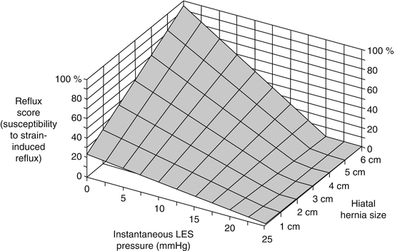
Fig. 2.2


Model of the relationship among lower esophageal sphincter pressure (x-axis), size of hernia (y-axis), and the susceptibility to gastroesophageal reflux induced by provocative maneuvers that increase abdominal pressure as reflected by the reflux score (z-axis). The statistical model was created by stepwise regression analysis of experimental data in which subjects performed these maneuvers while being monitored manometrically and fluoroscopically to detect reflux events. The overall equation for the model is as follows: reflux score = 22.64 + 12.05 (hernia size) − 0.83 (LES pressure) − 0.65 (LES pressure hernia size). The multiple correlation coefficient of this equation for the 50 subject data set was 0.86 (R 2 = .75) indicating that 75 % of the observed variance in susceptibility to stress reflux among individuals was accounted for by the size of hiatus hernia and the instantaneous value of LES pressure (From Sloan et al. [34], with permission). Going one step further, this same group revealed that the separation leads to greater propensity to reflux during abrupt increases in IGP. Reflux score is extremely low when either the LES is intact or the degree of axial displacement is limited. Thus, it appears that GERD requires at least two hits to the EGJ in order to occur
Stay updated, free articles. Join our Telegram channel
Full access? Get Clinical Tree
