Osteosarcoma
Presentation
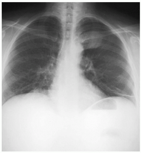
A 46-year-old woman is referred to you by an oncologist with complaints of leftsided back pain for the past several weeks. She has been followed by her oncologist for a history of lymphoma for which she has been previously treated. She denies any weight loss, fever, chills, or night sweats and is a nonsmoker. Her physical examination reveals tenderness over the region of the fourth and fifth ribs in the left paravertebral region. She presents with the following chest x-rays.
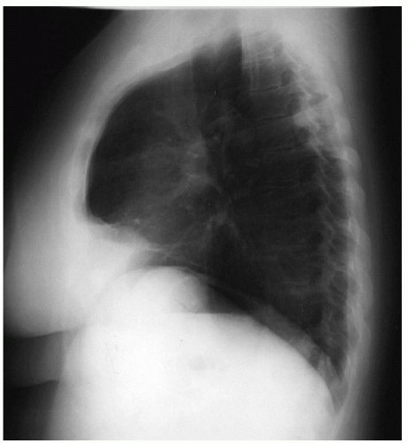
▪ Chest X-rays
Chest X-ray Report
There is a posterior, paravertebral chest wall mass at the level of the fourth and fifth ribs. The lung fields are clear without evidence of other masses. The mediastinum appears unremarkable, without evidence of masses or adenopathy. There are no pleural effusions.
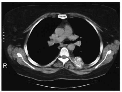
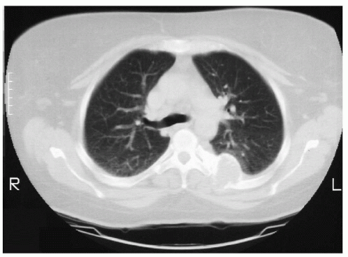
Computed tomography (CT) scans are performed to evaluate the chest x-ray finding.
▪ CT Scans
CT Scan Report
This study reveals a mass in the posterior paravertebral chest wall. The mass appears to be of bony origin with diffuse calcification within, and with adjacent rib destruction. There is underlying pleural thickening, with the mass protruding into the left hemithorax. There does not appear to be invasion into the lung parenchyma. There are no pleural effusions, lung nodules or masses, or mediastinal adenopathy.
Differential Diagnosis
The patient has a new symptomatic chest wall mass. The differential diagnosis of a chest wall mass in the adult includes both benign and malignant pathology. Chest wall tumors include neoplasms of bone or soft tissues and may be primary or metastatic disease. The incidence of malignancy for all chest wall neoplasms is about 50%. The malignant processes may be primary tumors of the chest wall or metastatic to the chest wall. Primary chest wall malignancies make up about half of the malignant pathology. The other half is composed of metastatic disease to the chest wall. The four most common metastatic lesions to the chest wall are from breast, renal, colon, and salivary primaries. Soft tissue tumors are the most common primary neoplasms of the chest wall. Malignant fibrous histiocytoma,
chondrosarcoma, and rhabdomyosarcoma are the most common malignant neoplasms of the chest wall. Cartilaginous tumors, desmoids, and fibrous dysplasia are the most common benign neoplasms of the chest wall. CT is needed to delineate the anatomy of the chest wall mass further and to plan subsequent therapy.
chondrosarcoma, and rhabdomyosarcoma are the most common malignant neoplasms of the chest wall. Cartilaginous tumors, desmoids, and fibrous dysplasia are the most common benign neoplasms of the chest wall. CT is needed to delineate the anatomy of the chest wall mass further and to plan subsequent therapy.
Stay updated, free articles. Join our Telegram channel

Full access? Get Clinical Tree


