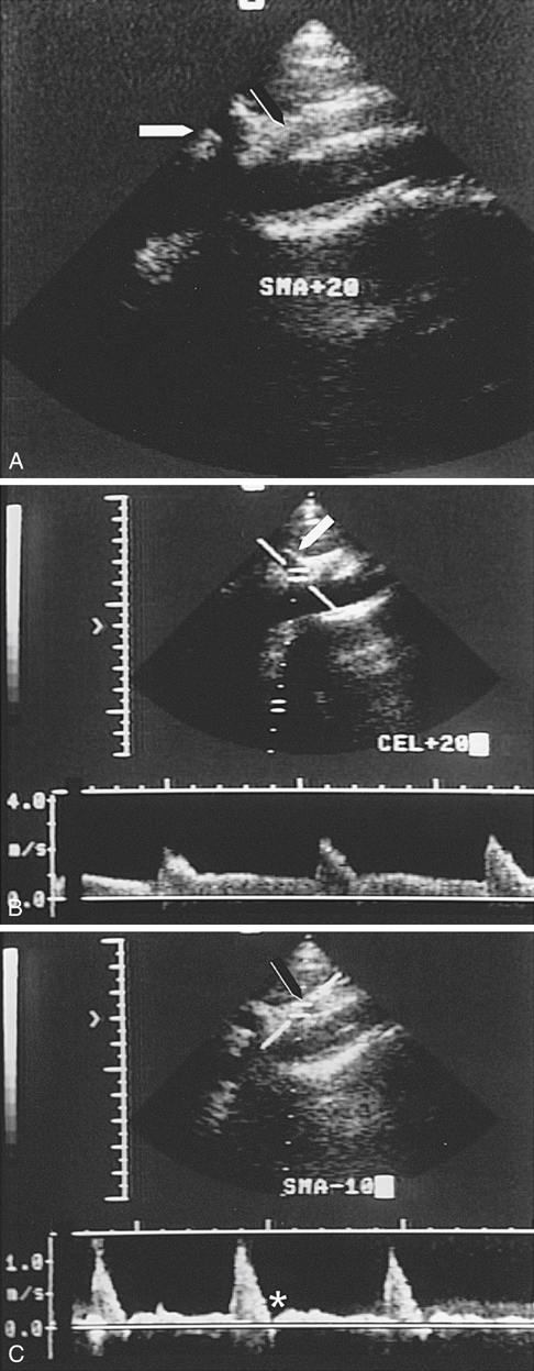
Duplex ultrasonography is an effective technique to provide accurate anatomic assessment of the vessels in the operating room. Duplex can be used experimentally to study splanchnic blood flow responses to various vasoactive agents. One approach is to use a small 7.5-MHz probe placed in a sterile sleeve accompanied by copious amounts of acoustic gel. The wound is filled with saline, and the surgeon places the probe directly over the area of reconstruction to be studied. Arteries under study are first identified at their origin along the aorta. Multiple transverse B-mode gray-scale images are obtained along the course of the reconstructed arteries, and any defects are imaged. Using longitudinal views, color flow images are then used to direct Doppler spectral analysis to insonate at the origins and endpoints of the endarterectomy, as well as to determine areas of increased or decreased blood flow velocity or turbulence (Figures 1 and 2).
Operative Evaluation of Renal and Visceral Arterial Reconstructions Using Duplex Sonography

Stay updated, free articles. Join our Telegram channel

Full access? Get Clinical Tree


