Open Transperitoneal Surgical Treatment of Nonruptured Infrarenal Aortic Aneurysms


Open Transperitoneal Surgical Treatment of Nonruptured Infrarenal Aortic Aneurysms


Yazan Duwayri and Gregorio A. Sicard


Open repair of an abdominal aortic aneurysm (AAA) had been the standard treatment for these lesions prior to the wide adoption of endovascular aneurysm repair (EVAR). Currently, in most developed countries, open repair of infrarenal AAAs is primarily performed in patients who do not fit the anatomic criteria for EVAR. In addition, some patients who may be good anatomic candidates for EVAR opt for open AAA repair if they are good surgical candidates and are unwilling or unable to participate in the necessary post-EVAR surveillance. Open repairs remain an integral part of every vascular surgery practice, with a transperitoneal approach being more widely used than the retroperitoneal approach, largely owing to surgeons’ familiarity with this incision. It is the optimal approach in patients with previous retroperitoneal surgery and when exposure of the right iliac vessels is needed.


Anatomic Preoperative Planning


Imaging by computed tomography angiography (CTA) or magnetic resonance angiography (MRA) should be done before any intervention for repair of AAA. Three-dimensional image reconstruction adds to the value of the images. Imaging delineates important anatomic characteristics of the aneurysm, including the shape, length, and calcifications of the aortic neck that can interfere with proximal clamp placement; the location and extent of intraluminal thrombus or plaque that can result in atheroembolism; iliac artery anatomy and size; and renal artery location. Patency of the celiac, superior mesenteric (SMA), and hypogastric arteries should also be assessed to determine the safety of intraoperative sacrifice of the inferior mesenteric artery (IMA). Important anatomic variations should also be sought and identified. These include venous anomalies such as left-sided or duplicated vena cava and retroaortic or circumaortic left renal vein. Congenital renal abnormalities are not uncommon and include horseshoe or pelvic kidney.


Technique


Exposure


The patient is placed in a supine position and prepared and draped from the nipples to the knees. A generous midline incision is made extending from the xiphoid process to the symphysis pubis. The abdominal contents are examined to rule out any undetected intra-abdominal pathology. If a concomitant pathology is found, although this is uncommon now, the overall guidelines are to treat the most life-threatening process first and avoid simultaneous operations. Typically, this means to proceed with AAA repair unless the unexpected findings carry increased risk for graft infection, such as an abscess or metastatic disease.


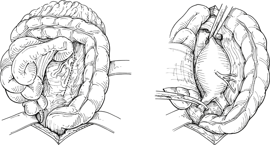
Following the abdominal exploration, the greater omentum and transverse colon are lifted superiorly and the small bowel is mobilized to the right side of the abdomen to expose the retroperitoneum. The retroperitoneum is then incised starting at the aortic bifurcation and moving superiorly, to the right of the inferior mesenteric vein, toward the ligament of Treitz to provide adequate exposure of the aortic neck region. The left renal vein can be visualized at this stage, and its absence should alert the surgeon to the possibility of a retroaortic position. The torrential hemorrhage from inadvertent injury of the anomalous left renal vein can be avoided by careful retraction and more careful placement of the aortic clamp (Figure 1).


Infrarenal AAAs treated in current times with open repair often have challenging necks. Owing to extensive thrombus or limited length of the aortic neck, both renal arteries may need to be identified and controlled before the infrarenal clamp is placed. In such cases, the left renal vein may need to be mobilized by division of its tributaries. If division of the left renal vein is found to be necessary in challenging cases, its gonadal and adrenal tributaries should be preserved.


The distal extent of exposure depends on the condition of both iliac arteries. Usually, control of the common iliac arteries just beyond their origin is sufficient. However, control of the hypogastric and external iliac arteries may be needed in the presence of iliac aneurysms. Both ureters should be clearly identified at this stage. Circumferential control of the iliac vessels should be avoided to prevent iliac vein injury. The splanchnic nerve plexus, important to sexual function, crosses the proximal aspect of the left common iliac artery and is also prone to injury during dissection in this location. To maintain the exposure during the remainder of the procedure, a self-retaining retractor is used.


Clamping


Before cross clamping, systemic heparinization is initiated with a dose approximating 80 U/kg. Additional heparin may be given at later stages to maintain activated clotting time at 250 to 300 seconds. Distal control is achieved first, to decrease the risk of distal atheroembolism. This is followed by proximal aortic clamping. Owing to the complex physiologic and humeral responses to aortic occlusion, this step must be clearly communicated to the anesthesiology team. Aortic clamping increases the systemic arterial pressure secondary to increased systemic vascular resistance. Similarly, it results in redistribution of blood from organs distal to the aortic clamp toward the central venous circulation. Although these changes are less severe with infrarenal clamping than with more proximal clamp locations, they can affect myocardial demand and can result in decompensation in patients with significant coronary artery disease or ventricular dysfunction. This can be minimized by normalizing preload, afterload, coronary blood flow, and contractility.

Stay updated, free articles. Join our Telegram channel

Aug 25, 2016 | Posted by in CARDIOLOGY | Comments Off on Open Transperitoneal Surgical Treatment of Nonruptured Infrarenal Aortic Aneurysms

Full access? Get Clinical Tree

Get Clinical Tree app for offline access