Open Surgical Treatment of Thoracoabdominal Aortic Aneurysms


Open Surgical Treatment of Thoracoabdominal Aortic Aneurysms


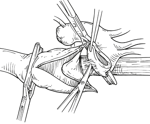
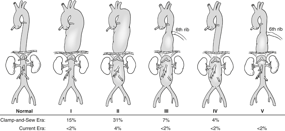
Kristofer M. Charlton-Ouw, Anthony L. Estrera and Hazim J. Safi


Aneurysms involving the thoracoabdominal aorta are uncommon, but they receive significant attention due to their complexity of repair and risk of end-organ impairment. Advances in preoperative planning, surgical technique, anesthesia, and postoperative management have led to improved outcomes, most notably with regard to postoperative neurologic deficit. Open repair of such aneurysms remains a formidable operation for both surgeon and patient. The pace of endovascular repair of thoracoabdominal aortic aneurysms is accelerating, but open repair remains the safest and most durable option for most patients.


Classification of Thoracoabdominal Aortic Aneurysms


The classification of thoracoabdominal aortic aneurysms is based on the extent of the involved aorta (Figure 1). Extent I is from the left subclavian to above the most proximal renal artery. Extent II is from the left subclavian artery to below the renal arteries. Extent III is from the sixth intercostal space to below the renal arteries. Extent IV is from T12 to below the renal arteries. Extent V, which was introduced in the last 2 decades, is from T6 to just above the renal arteries. The importance of the classification scheme is that it correlates with the incidence of neurologic deficits and mortality, especially when using the clamp-and-sew technique.


Clinical Manifestations


When symptoms are present before rupture, patients with thoracoabdominal aortic aneurysm complain of vague back or upper abdominal discomfort. Expanding aneurysms can cause pressure on adjacent structures, such as the lung, bronchus, esophagus, or recurrent laryngeal nerve. Axial imaging has largely replaced catheter-based aortography in the diagnostic assessment of thoracoabdominal aortic aneurysms. Computed tomography (CT) and magnetic resonance imaging are now commonplace and essential for assessing aneurysm morphology. Maximal aortic diameter, based on axial imaging or three-dimensional reconstructions, determines the need for operative intervention.


Thoracoabdominal aortic aneurysms can be expected to increase in size by 1 to 3 mm per year. Rupture is uncommon in aneurysms less than 4 cm in diameter. Rupture becomes unpredictable in aneurysms greater than 5 cm, but the risk continues to increase with size. The 5-year risk of rupture is greater than 30% for aneurysms larger than 6 cm. The goal of thoracoabdominal aortic aneurysm repair is prevention of rupture, because the mortality is greater than 90% in the first 24 hours after rupture.


We consider patients for repair of asymptomatic thoracoabdominal aortic aneurysm when maximal diameter is 5 to 6 cm. For patients at low risk of rupture, such as those with aneurysms less than 5 cm, we recommend repeat CT imaging at 6 months. Patients with connective tissue disorders, symptoms, saccular aneurysms, or diameter increases of more than 5 mm in 6 months are also at higher risk for rupture and are considered for earlier repair.


Once operative repair is indicated, preoperative assessment includes pulmonary function testing and echocardiography. Low preoperative left ventricular ejection fraction is a predictor for higher perioperative mortality. Additional testing for coronary artery disease is individualized. Optimal treatment of preoperative asymptomatic coronary artery disease remains controversial. Kidney function is evaluated by estimating the glomerular filtration rate (GFR). Values lower than 90 mL/min per 1.73 m2 body surface area indicate renal impairment. A low GFR is a powerful predictor of adverse outcome that is more sensitive than serum creatinine.


Open Surgical Technique


After anesthesia is induced and a double-lumen endotracheal tube is inserted, a pulmonary artery catheter, radial arterial line, and a cerebrospinal fluid (CSF) drain are placed. The patient is positioned in the right lateral decubitus position.


The thoracoabdominal incision is tailored to the extent of aneurysm but usually begins at the sixth rib and extends toward the midline. The latissimus dorsi is divided and the serratus anterior muscle is mobilized. The left lung is deflated and the chest is entered. The sixth rib is cut posteriorly or resected. Just inferior to the diaphragm, the retroperitoneal plane is entered and the viscera are rotated medially. The diaphragm is only partially transected in the muscular portion to avoid injuring the phrenic nerve.


Anticoagulation is started, and the inflow cannula is placed for distal aortic perfusion in the left inferior pulmonary vein. The outflow cannula is placed on the left common femoral artery using an end-to-side Dacron graft. The side-arm femoral cannulation technique prevents muscle ischemia and injury of the kidneys by allowing perfusion to the extremity. Distal aortic perfusion is initiated by withdrawing blood from the pulmonary vein and, using a roller pump, directing flow into the femoral cannula. When the aorta is clamped proximal to the aneurysm, flow is maintained to the viscera, renals, and lower extremities. A distal clamp is applied, and the aortic segment between the two clamps is opened (Figure 2). The aorta is separated from the esophagus, and the proximal anastomosis is sewn end to end with 3–0 polypropylene suture. The distal clamp is sequentially moved down the aorta to limit branch vessel ischemia.


The visceral aorta is repaired either as a patch anastomosis or as individual bypasses. We perform individual bypasses using a commercially available side-branched thoracoabdominal aortic graft (STAG). After the visceral vessels are attached to the graft, the proximal clamp is moved distally so that antegrade flow is restored. The timing of intercostal artery reattachment is guided by neuromonitoring changes. Loss of amplitude on motor-evoked or somatosensory-evoked potentials prompts us to reattach patent lower thoracic intercostal arteries as soon as feasible. Intercostal arteries are reattached as a patch to a side-arm graft. After the distal anastomosis is completed, the distal aortic perfusion pump is stopped. The graft is flushed and the clamps are released. The distal aortic perfusion cannulas are removed, and protamine is given to reverse the heparin anticoagulation. Chest tubes are placed, and the incision is closed in standard fashion.

Stay updated, free articles. Join our Telegram channel

Aug 25, 2016 | Posted by in CARDIOLOGY | Comments Off on Open Surgical Treatment of Thoracoabdominal Aortic Aneurysms

Full access? Get Clinical Tree

Get Clinical Tree app for offline access