Open Surgical Treatment of Ruptured Infrarenal Aortic Aneurysms


Open Surgical Treatment of Ruptured Infrarenal Aortic Aneurysms


James T. McPhee and Michael Belkin


Despite numerous medical and surgical technological advances in the past decade, rupture of an abdominal aortic aneurysm (AAA) still portends an exceedingly poor prognosis, with mortality rates greater than 90%, including patients who do not survive long enough to undergo medical evaluation. Reports from national data indicate that with the recent endovascular revolution, the annual number of patients experiencing ruptured AAA has slightly diminished as the numbers of elective endovascular aneurysm repair (EVAR) have increased nationally. This effect is more pronounced for men than for women, who continue to experience aneurysm rupture at an unchanged rate. To what extent newly supported AAA screening efforts for men older than 65 years could further decrease these rupture rates remains to be seen.


Diagnosis


The approach to the suspected AAA rupture has markedly changed in recent years as the availability of rapid diagnostic modalities has become more commonplace. The traditional surgical teaching that a patient with hypotension, back pain, and a pulsatile abdominal mass should be taken directly to the operating room still has value as a tenet; however, it is not universally applicable. Many centers now have a protocol-driven approach to suspected aneurysm rupture whereby a stance of permissive hypotension is maintained. This strategy is derived from the trauma literature and entails limiting volume resuscitation to the extent that the patient maintains consciousness and a detectable blood pressure to limit over-resuscitation and promote ongoing uncontrolled hemorrhage. If these tenets are met, many centers simultaneously ready the hybrid operating room for both open and endovascular repair while expeditiously performing a 64-slice computed tomography angiogram (CTA) of the abdomen and pelvis. Many trauma and referral centers can perform these studies within the emergency department and have three-dimensional reconstructions available to view in a matter of minutes.


In a patient who is truly hemodynamically unstable, only two things prevent the patient’s imminent demise: proximal control of the aorta and intravenous access to allow volume and blood-product resuscitation. With this scenario, it is appropriate to follow the time-tested principle of rapid transit to the operating room, pausing only to send a blood sample to the blood bank with the intent of having six units of blood immediately available, as well as having other products including fresh frozen plasma (FFP), platelets, and cryoprecipitate sent to the operating room to limit the unavoidable dilutional coagulopathy that will ensue.


In patients who are systemically anticoagulated on warfarin for other medical reasons, anticoagulation should be rapidly reversed. This reversal is traditionally performed with a combination of vitamin K and FFP. Anticoagulation should not delay the transfer process. Newer agents are available, such as prothrombin complex concentrates (PCCs) and recombinant activated factor VII (rFVIIa), that can allow a rapid reversal of warfarin-induced coagulopathy with a small volume of fluid. Enthusiasm for the ease and rapidity of the administration of these products must be tempered by the unmeasured potential for thrombotic complications, as well as the exorbitant health care costs associated with their use. The cell-saver autotransfusion device should be used if it is available because it can decrease the requirement for banked blood to some degree.


Procedure


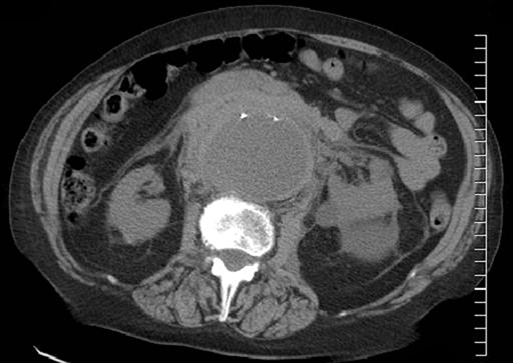
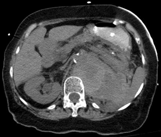
Upon arrival in the prewarmed operating room, a Foley catheter is placed, and an adequate patient warming device (Arctic Sun, Medivance, Inc, Louisville, CO) is placed. Simultaneous wide preparation of the patient, inclusive of the nipples to below the knees, is performed while intravenous and arterial access are obtained. Advanced central venous access, pulmonary artery catheters, and echocardiography probe placement should be deferred until the surgery is begun and may be done simultaneously by the anesthesia team as proximal control of the aorta is obtained. Default patient positioning is supine because more than 95% of AAAs are infrarenal and may be approached transabdominally (Figure 1). If preoperative imaging was performed and a perivisceral or thoracoabdominal component exists (Figure 2), right lateral decubitus positioning allows easier access to the entire thoracoabdominal aorta via a retroperitoneal approach.


Constant constructive communication with the anesthesia team will allow the most efficient forward motion through this preparatory phase, which usually has the greatest likelihood of delaying progress. After the patient is fully prepped and draped, general anesthesia is induced. If induction of anesthesia leads to loss of vital signs, the surgical team must move expeditiously to gain control of the proximal aorta to allow the resuscitative effort to succeed. This may be achieved either with a rapid and generous midline incision and manual occlusion of the supraceliac aorta or, less commonly, with a left anterior thoracotomy if the patient’s body habitus or prior abdominal surgery preclude rapid intraabdominal access. More recently, internal clamping of the aorta has been described by which a hybrid approach is used to endovascularly place an aortic occlusion balloon above the level of rupture if femoral or brachial arterial access is readily obtained in a room with angiographic capabilities. This endovascular technique may be useful even in cases where open repair is planned because it avoids the severe hypotension associated with loss of an abdominal compartment tamponade.


Preoperative imaging can help steer the focus of the dissection to determine optimal clamp placement. If preoperative imaging or initial exploration confirm a true infrarenal rupture with an adequate infrarenal neck, it might not be necessary to establish supraceliac control. In this case, the dissection proceeds in the same manner as for elective repair through a generous midline laparotomy (detailed later).


Some surgeons prefer to routinely obtain supraceliac proximal control. This practice may be of additional benefit when the aortic anatomy is completely unknown. This exposure is accomplished by mobilizing the left lateral hepatic segment of the liver to the patient’s right with a combination of blunt dissection and electrocautery. The esophagogastric junction is identified by palpation of the nasogastric tube or transesophageal echo probe and is gently distracted to the patient’s left side.


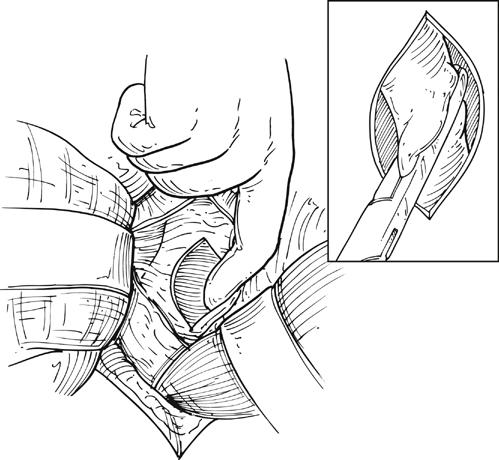
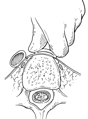
The gastrohepatic ligament is divided after palpation for a replaced or accessory left hepatic artery, which is present in 10% to 20% of patients and courses in a transverse orientation toward the left hepatic lobe. The safest practice is to directly dissect down onto the aortic pulse. A combination of sharp and blunt dissection is used to splay the fibers of the diaphragmatic crus and retroperitoneal tissues until the white sheen of the aortic wall is revealed. Gentle blunt finger dissection is used directly on the aorta laterally and medially to develop enough space to insert a deep aortic clamp into place (Figure 3). If at any point during this dissection the aortic pulse is lost, immediate aortic control may be obtained by direct digital compression of the aorta posteriorly onto the vertebral column (Figure 4).


Stay updated, free articles. Join our Telegram channel

Aug 25, 2016 | Posted by in CARDIOLOGY | Comments Off on Open Surgical Treatment of Ruptured Infrarenal Aortic Aneurysms

Full access? Get Clinical Tree

Get Clinical Tree app for offline access