Open Surgical Treatment of Popliteal Artery Aneurysms


Open Surgical Treatment of Popliteal Artery Aneurysms


Jose L. Trani and Jeffrey P. Carpenter


Popliteal artery aneurysms (PAAs) are uncommon, having an estimated incidence in the general population of 0.1%. They are the third most common aneurysmal pathology encountered in clinical practice, ranking behind both aortic and cerebrovascular aneurysms. Half to three quarters of patients with PAAs come to the hospital with bilateral lesions. Patients with PAAs have a 30% to 50% chance of a concomitant abdominal aortic aneurysm (AAA), the latter being more likely in patients with bilateral aneurysms. The converse statement, that a PAA is present in a patient with aortic aneurysms, is true in 3% to 12% of patients.


Diagnosis of a PAA mandates lifelong surveillance. It is estimated that half to three quarters of those with these aneurysms either come to the hospital with synchronous aneurysms or develop a separate aneurysm at a distant location within 10 years, mandating continued surveillance after the PAA is addressed.


Aneurysm formation is typically defined as a greater than 50% increase in diameter relative to the accepted normal size of the artery. The accepted diameter of a normal popliteal artery is between 0.7 and 1.1 cm. PAAs have growth rates between 0.7 and 1.5 mm yearly, and larger aneurysms expand between 1.5 and 3.5 mm yearly. The timing of intervention for asymptomatic aneurysms is somewhat controversial given the lack of prospective randomized data on which to base management decisions.


PAAs were described as early as the 3rd century AD. The earliest strategies for treatment involved inducing thrombosis of the vessel. Various techniques, including knee flexion for prolonged periods, compression bandaging, and direct manual compression, were undertaken in attempts to induce thrombosis. These maneuvers were maintained, on average, for 2 weeks. In the 18th century, John Hunter carried out a surgical intervention consisting of proximal ligation of the aneurysm and reliance on geniculate collaterals to maintain perfusion to the leg.


Reconstructive endoaneurysmorrhaphy, whereby the aneurysm was treated by suturing the walls of the aneurysm together over a catheter, thus maintaining in-line flow to the foot, was described in 1906. This time period also saw the use of interposition grafting using either popliteal or greater saphenous vein as conduit. Exclusion and vein bypass was introduced by Edwards in 1969. Exclusion and bypass remains the most common form of treatment in current surgical practice.


Clinical Presentation


The most limb-threatening presentation of a patient with a PAA is that of acute limb ischemia. This is typically caused by thrombosis of the popliteal aneurysm, with concomitant loss of the runoff vessels through a combination of chronic distal embolization and acute thrombosis of the remaining vessel. This presentation carries with it the worst prognosis with respect to limb salvage and necessitates urgent surgical intervention. From 40% to greater than 50% of all patients in published series present in this manner, a number that has remained distressingly high with little change since the 1980s.


Despite the severity of the initial presentation, limb-salvage rates have remained good. A recent meta-analysis demonstrated 30-day amputation rates of 14.1% in patients presenting acutely, with approximately 20% of the total number of amputations performed as a primary procedure without thrombectomy, thrombolysis, or bypass. Smaller series from other centers have reported higher amputation rates, exceeding a third of patients treated. Rupture as the initial presentation is a relatively uncommon acute presentation occurring in only 2% of patients. Fortunately, in this setting, life-threatening hemorrhage is rare because of the constraints of the popliteal fossa. Although it is not life threatening, a ruptured PAA is limb threatening, with reported amputation rates of 50% to 75%.


A second group of patients come to the hospital with symptoms of arterial insufficiency, ranging from claudication to tissue loss. This group represents up to 40% of all patients with PAAs. Repair of these aneurysms in this subgroup of patients should be undertaken regardless of their size given that distal embolization has already begun to compromise the popliteal artery runoff vessels. In addition, this approach provides better outcomes with respect to graft patency, limb loss, and mortality.


A much smaller group of patients with PAAs have symptoms caused by physical compression of venous or nerve structures surrounding the aneurysm within the popliteal fossa. This occurs almost exclusively in patients with aneurysms that are greater than 3 cm. Surgery for these patients decompresses the aneurysm to relieve symptoms and excludes the aneurysm.


In most published series, asymptomatic patients account for between 25% and 50% of patients who present with a PAA. These patients develop symptoms at a rate of approximately 14% per year. Up to 40% of this group have absent pedal pulses, suggesting the presence of distal embolization even in the absence of any clinical manifestations. When Dawson and colleagues compared asymptomatic patients with and without pulses they found that 34% of patients with pulses developed symptoms within 3 years compared to 86% of those without pulses at their initial presentation.


Diagnosis and Imaging


Asymptomatic PAAs may be detected by physical examination. Given the relatively common concomitance of AAAs and PAAs, patients with aortic aneurysms merit an evaluation of their popliteal vessels in the office. Unfortunately, there is a relatively poor correlation with positive and negative predictive values in detecting PAAs, with just over 50% identified through an abnormal palpable pulse. Ultrasound provides a safe, noninvasive, office-based means of identifying PAAs and distinguishing them from other popliteal fossa entities. Through the use of color flow in conjunction with B-mode imaging, the aneurysm’s diameter and the presence or absence of thrombus can be determined. Duplex ultrasound also offers the ability to evaluate the patency of the tibial and peroneal vessels. Limitations of ultrasonography include operator dependence and difficulty correlating location and extent of disease to the surrounding anatomic structures. However, ultrasonography is the recommended modality for following small aneurysms.


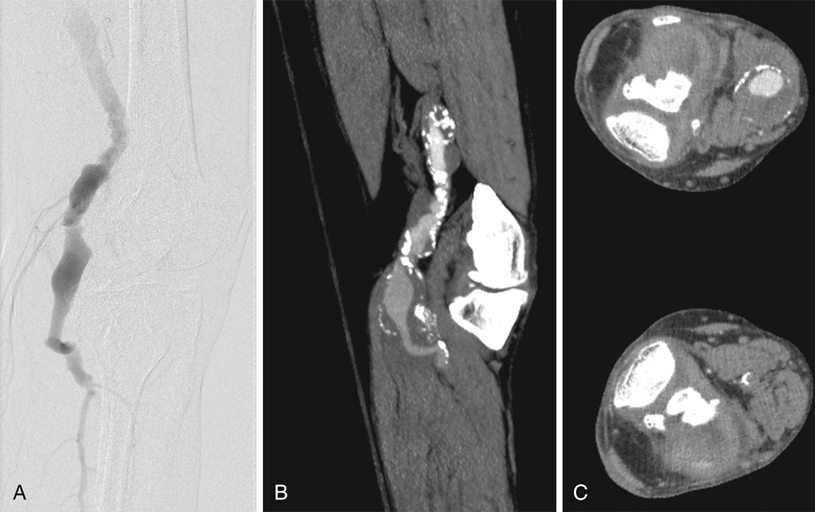
Computed tomography angiography (CTA) and magnetic resonance angiography (MRA) offer noninvasive ways to obtain information regarding the extent of the aneurysm and the status of runoff vessels. Images obtained through these modalities can be used to determine the best surgical approach for repair. CTA is especially useful if consideration is to be given to an endovascular repair. Conventional angiography provides only information about the internal diameter of a vessel and thus can underestimate the diameter of a PAA containing thrombus (Figure 1). The principal role of conventional angiography lies in its use for evaluating runoff vessels, particularly in the acute setting, and in initiating and follow-up imaging of thrombolysis.


Surgical Management


All patients who come to the hospital with symptoms should be offered repair. In addition, most authors would recommend intervention in asymptomatic patients once the diameter reaches 2.0 or 2.5 cm, citing the eventual likelihood of progression to symptoms and the better limb-preservation outcomes with lower risk of perioperative complications. Poor-risk surgical candidates should be counseled about other options, such as endovascular repair or surveillance. One small cohort of seven nonoperative patients with aneurysms greater than 3 cm was followed prospectively, and all became symptomatic within 30 months, with three experiencing thrombosis of their aneurysm.


Open operative intervention in the elective setting can be performed through either a medial or a posterior approach. The medial approach is the technique more familiar to most surgeons, and this is the approach currently most often chosen for repair. Separate surgical incisions are made above and below the knee on the medial aspect of the thigh and the leg to expose normal artery above and below the aneurysm. Should the pathology extend beyond the popliteal artery, this approach can easily facilitate either more proximal or more distal arterial access than the posterior approach. This is the technique of choice if preoperative imaging demonstrates a need to use a tibial vessel for the distal anastomosis. A medial approach also facilitates the use of the greater saphenous vein for a conduit. Following successful bypass around the PAA, the native vessel should be ligated distal to the proximal anastomosis and proximal to the distal anastomosis to prevent continued flow through the aneurysm and the concomitant risks of expansion and distal embolization. Aneurysm decompression through this approach might be difficult and can require division and repair of the medial head of the gastrocnemius muscle.


The posterior approach is performed by making an S-shaped incision through the popliteal fossa with the patient in the prone position. Proximally, the incision is made on the medial aspect of the thigh, with the transverse portion of the incision made before the knee crease. Care must be taken to not extend the longitudinal portion of the inferiolateral incision too distally because this increases the risk of peroneal nerve neuropraxia caused by a retraction injury. The proximal popliteal artery is exposed by separating the semimembranosus and semitendinosus muscles medially from the long head of the biceps femoris laterally. The distal popliteal artery is exposed by separating the two heads of the gastrocnemius muscle. The deep fascia should be incised longitudinally to avoid injury to the median cutaneous sural nerve that travels with the small saphenous vein. In the proximal portion of the incision, the tibial nerve is the most superficial major structure encountered. The popliteal artery and vein is deeper in the popliteal fossa with the vein lateral to the artery. Advantages of a posterior approach include easy appreciation of anatomic structures, including proximal and distal extent of the PAA, and the ability to decompress the aneurysm in patients with compressive symptoms.


Options for repair using this approach include either a bypass graft with interval ligation or an interposition graft similar to that used for open infrarenal aneurysm repair. An interposition graft offers the advantage of identification and oversewing of geniculate collaterals that can continue to pressurize the aneurysm sac after a bypass and ligation. The primary limitations of this approach are limited opportunities for additional exposure either proximally or distally should pathology necessitate. Additionally, vein conduit can be difficult to harvest from a patient in the prone position. Solutions include beginning the procedure with the patient supine to procure greater saphenous vein then moving the patient to the prone position or using the lesser saphenous vein, which can be obtained with the patient in the prone position.


An analysis of the Swedvasc registry compared 1-year results between medial and posterior approaches. The medial approach was used just over 10 times more often than the posterior approach. Neither 30-day (93.0% vs. 93.9%) nor 1-year (87.0% vs. 90.3%) patency rates were statistically different between the posterior and the medial approaches, respectively (Table 1). The authors also found that these two approaches also had similarly low (3.7% vs. 2.6%) late amputation rates resulting from either of the two procedures. Some authors have offered dissenting opinions, with single-center data demonstrating superior patency and limb salvage with the posterior approach.


Aug 25, 2016 | Posted by in CARDIOLOGY | Comments Off on Open Surgical Treatment of Popliteal Artery Aneurysms

Full access? Get Clinical Tree

Get Clinical Tree app for offline access