Open Surgical and Endovascular Management of Ruptured Abdominal Aortic Aneurysm

Chapter 41 Open Surgical and Endovascular Management of Ruptured Abdominal Aortic Aneurysm


A ruptured abdominal aortic aneurysm (AAA) is a relatively common but catastrophic problem that, if untreated, will almost always result in the death of a patient. With treatment, which can consist of open surgical repair or endovascular graft exclusion of the ruptured AAA, many patients will survive and live out their normal lifespan. This chapter will discuss important aspects of the open surgical and endovascular graft repair of ruptured AAAs that have a rupture site in the aorta or common iliac arteries. Because the preoperative, intraoperative, and postoperative management associated with open surgical repair is well known and well standardized, this chapter will stress only crucial and less well-known aspects of these areas of open repair. The remainder of this chapter will then address the history and current status of endovascular treatment of ruptured AAAs, with an emphasis on endovascular aneurysm repair (EVAR) by endograft exclusion of the ruptured AAA. The text will also emphasize key management strategies, technical maneuvers, and adjuncts that are important in achieving optimal outcomes in the very sick patient group with a ruptured AAA.


Open Surgical Management: Key Points


Crucial Diagnostic Triad


Pain, syncope, and a known or palpable AAA are a crucial diagnostic triad. If a patient has all three elements, he or she should be considered to have a presumptive diagnosis of a ruptured AAA. If open surgical treatment is planned, the patient should be taken immediately to the operating room and prepared for operation. No further diagnostic evaluation is necessary. Although an occasional patient will have another catastrophic problem, most of these patients require a laparotomy anyway. The only exceptions are an acute myocardial infarction and pancreatitis. The former can be suggested because of electrocardiogram findings, the latter because of a high hematocrit and an elevated amylase.


The history of syncope is usually a manifestation of hypovolemia if cardiac causes can be excluded. The known AAA may be confirmed by history or the presence of a palpable pulsatile mass on physical examination. Although the pain element of the triad is usually in the mid-abdomen or back, it may be in the chest, lower abdomen, flank, or groin. These unusual locations are caused by the pressure of the hematoma, which might not be centrally located and therefore produce unusual pain patterns. One example would be flank and groin pain mimicking a renal or ureteral stone. Another example would be left lower quadrant pain and a mass mimicking diverticulitis.


If a patient only has two elements of the triad, further emergent diagnostic studies with ultrasound or CT scanning are justified. However, a member of the surgical team should be with the patient during these urgent studies in case hemodynamic collapse occurs. The main purpose of these studies is usually just to confirm the presence of an AAA, although occasionally blood can be seen outside the wall of the AAA or other important information can be provided.


Strategies in Planning Operative Treatment


When the diagnosis of a ruptured AAA is presumed (i.e., three elements of the triad) or confirmed if two elements are present initially, a decision can be made to proceed with operative treatment. Although the patient should be taken immediately to the operating room, other elements or strategies in the treatment are important. The team must be assembled; this includes junior and senior surgeons, nurses, and anesthesiologists. Because these ruptured AAA emergencies often occur at night or on weekends, assembling the team needed to treat them can take some time and organization. However, most operative procedures should not begin and the patient should not be anesthetized until all members of the team are present. While the team is gathering, lines and nasogastric and bladder tubes can be placed, instruments set up, and the abdomen prepared and draped. Only when all personnel are present should the patient be put to sleep, because circulatory collapse is prone to occur when the anesthesia removes the sympathetic compensation that maintains the circulation in the hypovolemic state. Only if the patient’s circulation collapses completely should a junior member of the team begin the operation before the whole team is fully assembled and operative preparations are completed. The strategic principle should be to get the patient rapidly to the operating room, but then proceed deliberately and only anesthetize the patient and begin the operation when everyone and everything is set and in place.


One final strategy that is as effective in open repair management, as it is in endovascular treatment, is the use of hypotensive hemostasis or restricted fluid resuscitation. This strategy curtails bleeding by allowing the patient to be hypotensive throughout the preoperative and intraoperative phases of treatment. This subject will be discussed in greater detail in the endovascular treatment section.


Key Technical Points for Open Repair of Ruptured Abdominal Aortic Aneurysms


Once the abdomen is opened through a short midline incision, the base of the mesentery is inspected; if blood is seen, the diagnosis of rupture is confirmed. The incision is then extended from xiphoid to pubis.


If the area of the infrarenal neck of the AAA is involved with hematoma, supraceliac aortic control should be obtained as shown in Figure 41-1; this is done without direct vision by tearing the lesser omentum with a finger (see Figure 41-1A). This process exposes the retroperitoneum above the celiac axis and pancreas (see Figure 41-1B). Next, using the gloved index finger and its nail, the posterior peritoneum over the diaphragmatic crura is torn as indicated by the line in Figure 41-1C, and the fingertip is pushed through the muscle fibers until the periadventitia of the aorta is encountered (see Figure 41-1D). Next, the finger bluntly dissects medially and laterally in the same plane, creating a potential space on both sides of the aorta (see Figure 41-1E to G). Two fingers are placed in the potential space and pulled caudally (see Figure 41-1G). A large curved DeBakey clamp is then placed vertically above the fingers into the space to occlude the aorta (see Figure 41-1H). The clamp will be occlusive only if the aorta is freed from the crural fibers anteriorly, medially, and laterally. It is also crucial to have a nasogastric tube in the esophagus during these maneuvers so that this structure can be identified and protected from injury.
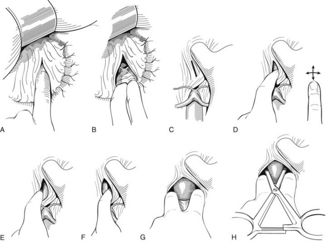


Following placement of this clamp, it is essential to minimize the time of supraceliac aortic occlusion (15 to 20 minutes is tolerable) to minimize liver and gut ischemia. To do this, the anterior wall of the AAA is exposed in its most prominent portion away from all venous and other structures. The anterior wall of the AAA is incised, and a finger is placed within the AAA sac to identify the aortic neck. This may be deviated sharply to the right, left or anteriorly because of aortic tortuosity associated with AAAs. With a finger inside to identify the position of the neck, it can gently be dissected bluntly from adjacent structure in the periadventitial plane without injuring major veins. Once dissected anteriorly, medially, and laterally, an infrarenal aortic clamp can be placed and the suprarenal clamp is removed.


The iliac arteries can then be identified from without and clamped with gently placed atraumatic clamps. If they cannot be easily identified by palpation from without, they can be identified from within the lumen by a finger within the AAA sac. Clamps can then be placed on them without further dissection or with minimal dissection.


Clot is then removed from the interior of the AAA. Bleeding lumbar and any other branches are identified and controlled with sutures from within. A prosthetic graft is then sutured in place from within the lumen of the AAA. The posterior walls of the aortic and iliac arteries should never be dissected to prevent damage to other vascular structures. The posterior walls of the aorta and iliac arteries should not be transected to avoid injuries to adherent major veins and minimize bleeding from other vascular structures.


Although controversial, heparin should be administered as soon as aortic clamp control is obtained. All dissection should be minimized in these ruptured AAA repairs to minimize injury to other structures and blood vessels, the position of which can be distorted by the AAA and the hematoma. Otherwise, the repair should be as much like a standard AAA repair as possible. Care should be taken to monitor and treat hypothermia and coagulation defects. Lastly, care should be taken to diagnose and treat abdominal compartment syndrome as detailed in the endovascular section that follows.


Stay updated, free articles. Join our Telegram channel

Jul 1, 2016 | Posted by in CARDIOLOGY | Comments Off on Open Surgical and Endovascular Management of Ruptured Abdominal Aortic Aneurysm

Full access? Get Clinical Tree

Get Clinical Tree app for offline access